Copyright
©2008 The WJG Press and Baishideng.
World J Gastroenterol. Jun 7, 2008; 14(21): 3338-3349
Published online Jun 7, 2008. doi: 10.3748/wjg.14.3338
Published online Jun 7, 2008. doi: 10.3748/wjg.14.3338
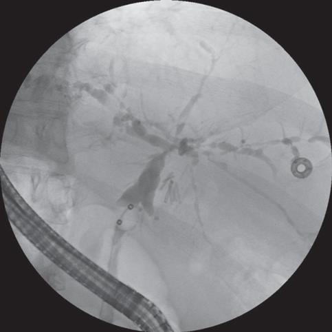
Figure 1 Cholangiographic finding in PSC.
Cholangiogram demonstrating multifocal strictures with intervening saccular dilatation of both intrahepatic and extrahepatic bile duct characteristic of PSC. (Photograph courtesy of Dr. Rahul Pannala).
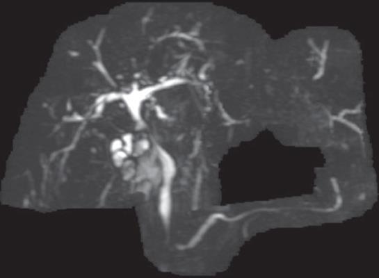
Figure 2 Cholangiographic findings in PSC.
Magnetic resonance cholangiography demonstrating findings of PSC.
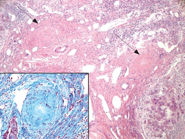
Figure 3 Fibro-obliterative lesions in PSC.
Image shows an expanded portal area without two distinct fibro-obliterative lesions (arrows) in end-stage primary sclerosing cholangitis. There is no intact bile duct present in this portal area, only cross-sections of portal vein and hepatic artery branches (H&E, original magnification, 100 ×). Inset: Higher magnification of the fibro-obliterative lesion (Masson trichrome, × 400). (Photograph courtesy of Dr. Schuyler Sanderson).
- Citation: Silveira MG, Lindor KD. Clinical features and management of primary sclerosing cholangitis. World J Gastroenterol 2008; 14(21): 3338-3349
- URL: https://www.wjgnet.com/1007-9327/full/v14/i21/3338.htm
- DOI: https://dx.doi.org/10.3748/wjg.14.3338


