Copyright
©2008 The WJG Press and Baishideng.
World J Gastroenterol. Apr 21, 2008; 14(15): 2448-2450
Published online Apr 21, 2008. doi: 10.3748/wjg.14.2448
Published online Apr 21, 2008. doi: 10.3748/wjg.14.2448
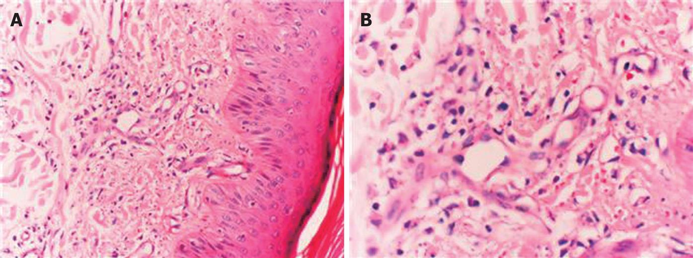
Figure 1 Skin biopsy specimen (HE, × 200) showing polymor-phonuclear cells and lymphocyte infiltration in and around the vessels of dermis beneath the multilayered keratinized squamous epithelium with some nuclear debris in the interstitium (A) and fibrin deposits in the vessel wall and nuclear debris (B).
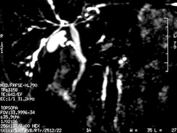
Figure 2 Magnetic resonance cholangiopancreaticography demonstrating ductal irregularities and beading appearence in the distal branches of the right and left hepatic canals.
- Citation: Akbulut S, Ozaslan E, Topal F, Albayrak L, Kayhan B, Efe C. Ulcerative colitis presenting as leukocytoclastic vasculitis of skin. World J Gastroenterol 2008; 14(15): 2448-2450
- URL: https://www.wjgnet.com/1007-9327/full/v14/i15/2448.htm
- DOI: https://dx.doi.org/10.3748/wjg.14.2448


