Copyright
©2007 Baishideng Publishing Group Co.
World J Gastroenterol. Aug 21, 2007; 13(31): 4278-4281
Published online Aug 21, 2007. doi: 10.3748/wjg.v13.i31.4278
Published online Aug 21, 2007. doi: 10.3748/wjg.v13.i31.4278
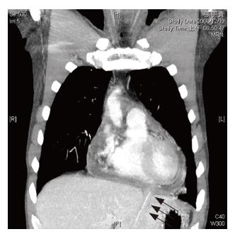
Figure 1 High resolution chest computed tomography (CT) revealed a moderate amount of pericardial effusion with possible superimposed infection.
Thickness of pericardium and left lobe liver abscess were found. A straight tubular structure about 6 cm in length transverses the lateral segment of liver to pericardial space and unknown foreign body was suspected.
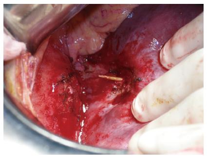
Figure 2 Operative findings revealed severe adhesion between liver and diaphragm.
After the space was divided between the liver and diaphragm, a toothpick was found through the liver into pericardium.
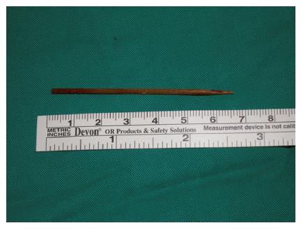
Figure 3 Gross findings of a toothpick measuring 6.
5 cm in length.
- Citation: Liu YY, Tseng JH, Yeh CN, Fang JT, Lee HL, Jan YY. Correct diagnosis and successful treatment for pericardial effusion due to toothpick injury: A case report and literature review. World J Gastroenterol 2007; 13(31): 4278-4281
- URL: https://www.wjgnet.com/1007-9327/full/v13/i31/4278.htm
- DOI: https://dx.doi.org/10.3748/wjg.v13.i31.4278


