Copyright
©2007 Baishideng Publishing Group Co.
World J Gastroenterol. Aug 21, 2007; 13(31): 4274-4277
Published online Aug 21, 2007. doi: 10.3748/wjg.v13.i31.4274
Published online Aug 21, 2007. doi: 10.3748/wjg.v13.i31.4274
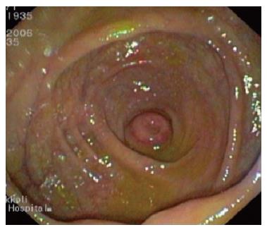
Figure 1 Endoscopic view of cecum and intussuscepted appendix.
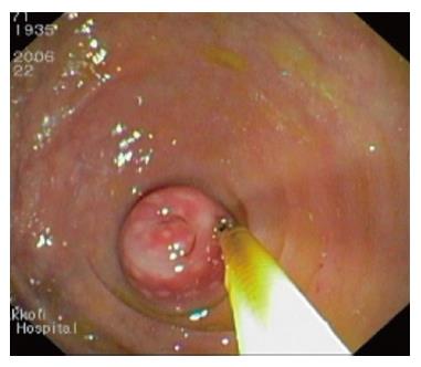
Figure 2 Colonoscopic finding: sessile polypoid lesion touched by closed pence.
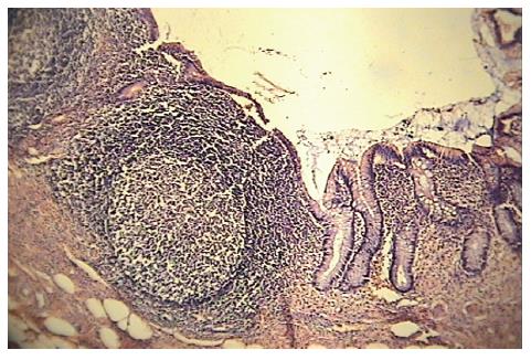
Figure 3 The lamina propria of the appendix is obliterated by lymphoid aggregates with prominent germinal centers.
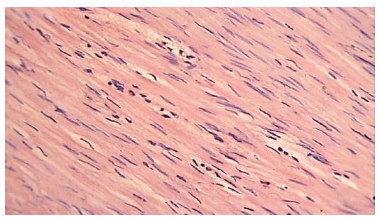
Figure 4 Muscularis propria is infiltrated by few polymorphonuclear leukocytes.
- Citation: Tavakkoli H, Sadrkabir SM, Mahzouni P. Colonoscopic diagnosis of appendiceal intussusception in a patient with intermittent abdominal pain: A case report. World J Gastroenterol 2007; 13(31): 4274-4277
- URL: https://www.wjgnet.com/1007-9327/full/v13/i31/4274.htm
- DOI: https://dx.doi.org/10.3748/wjg.v13.i31.4274


