Copyright
©2007 Baishideng Publishing Group Co.
World J Gastroenterol. Aug 21, 2007; 13(31): 4270-4273
Published online Aug 21, 2007. doi: 10.3748/wjg.v13.i31.4270
Published online Aug 21, 2007. doi: 10.3748/wjg.v13.i31.4270
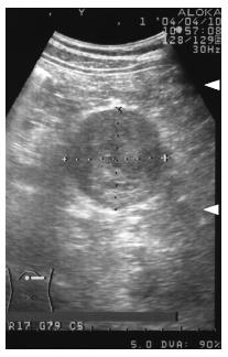
Figure 1 Abdominal ultrasound examination, showing an encapsulated, rather heterogeneous, hypoechoic tumor, 6.
5 cm in maximum diameter, with a beak sign.
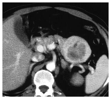
Figure 2 Helical dynamic CT, revealing an irregularly enhanced tumor with pooling of contrast medium in the delayed phase.
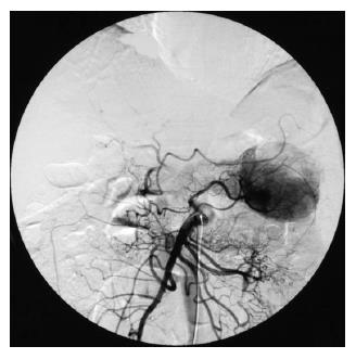
Figure 3 Abdominal angiography.
The tumor was hypervascular, fed by both the splenic artery and dorsal pancreatic artery from the superior mesenteric artery.
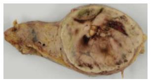
Figure 4 Macroscopic findings.
The cut surface of the mass was reddish-yellow, with focal hemorrhage and a necrotic appearance.
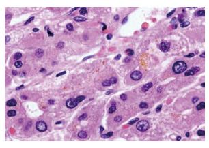
Figure 5 Histological findings (HE, x 400).
The tumor cells grew in trabeculae that were separated by sinusoid-like blood spaces. These tumor cells were polygonal with fine granular eosinophic cytoplasm, resembling hepatocytes.
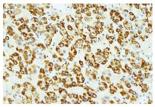
Figure 6 Immunohistochemical staining (HEPPAR-1).
The tumor cells were positive for HEPPAR-1, suggesting that they had the nature of hepatocytes.
- Citation: Kubota K, Kita J, Rokkaku K, Iwasaki Y, Sawada T, Imura J, Fujimori T. Ectopic hepatocellular carcinoma arising from pancreas: A case report and review of the literature. World J Gastroenterol 2007; 13(31): 4270-4273
- URL: https://www.wjgnet.com/1007-9327/full/v13/i31/4270.htm
- DOI: https://dx.doi.org/10.3748/wjg.v13.i31.4270


