Copyright
©2007 Baishideng Publishing Group Co.
World J Gastroenterol. May 7, 2007; 13(17): 2479-2483
Published online May 7, 2007. doi: 10.3748/wjg.v13.i17.2479
Published online May 7, 2007. doi: 10.3748/wjg.v13.i17.2479
Figure 1 Tumor volumes at different time-points in Hepal-6 (A) and H22 (B) models.
Cont-Ab: mice transfused with control antibodies; Cont-NS: mice transfused with NS; anti-mAb: mice transfused with anti-endoglin mAb. aP < 0.05, bP < 0.01. Data are shown as mean ± SD, n = 10 in each group.
Figure 2 The survival rates at different time-points in Hepal-6 (A) and H22 (B) models.
Log-rank test, P < 0.01, n = 10 in each group.
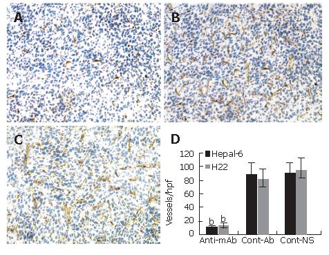
Figure 3 Significant decrease in microvessel density (stained as purple color) in the tumor tissues from the mice transfused with anti-endoglin mAb (A) compared to the mice transfused with control antibodies (B) and the mice transfused with NS (C).
Quantitative analysis of microvessels in Hepal-6 and H22 models (D), the bars represent mean ± SD, bP < 0.001.
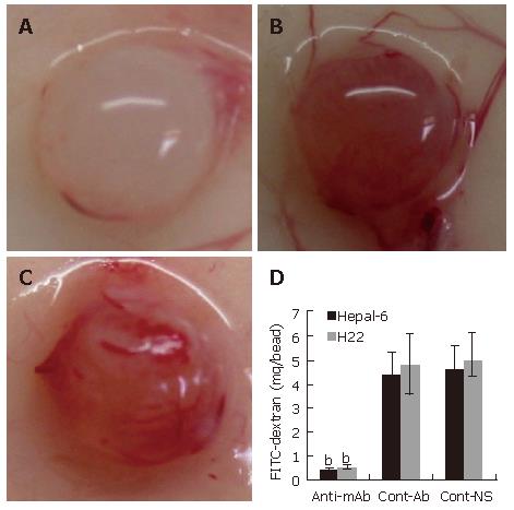
Figure 4 Significant inhibition of angiogenesis in the tumor cell-encapsulated beads from the mice transfused with anti-endoglin mAb (A) than that in the tumor tissues from the mice transfused with control antibodies (B) and the mice transfused with NS (C).
Quantitative determination of FITC-dextran uptake in Hepal-6 and H22 models (D); the bars represent mean ± SD, bP < 0.001.
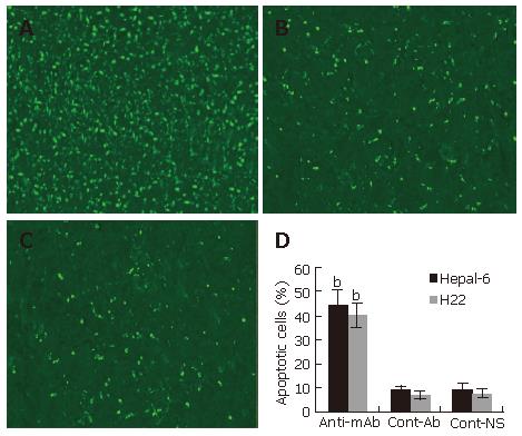
Figure 5 Significantly increased apoptosis in the tumor tissues from the mice transfused with anti-endoglin mAb (A) compared to the mice transfused with control antibodies (B) and the mice transfused with NS (C).
Quantitative analysis of apoptotic cells in Hepal-6 and H22 models (D), the bars represent mean ± SD, bP < 0.001.
- Citation: Tan GH, Huang FY, Wang H, Huang YH, Lin YY, Li YN. Immunotherapy of hepatoma with a monoclonal antibody against murine endoglin. World J Gastroenterol 2007; 13(17): 2479-2483
- URL: https://www.wjgnet.com/1007-9327/full/v13/i17/2479.htm
- DOI: https://dx.doi.org/10.3748/wjg.v13.i17.2479


