Copyright
©2007 Baishideng Publishing Group Co.
World J Gastroenterol. Mar 14, 2007; 13(10): 1622-1625
Published online Mar 14, 2007. doi: 10.3748/wjg.v13.i10.1622
Published online Mar 14, 2007. doi: 10.3748/wjg.v13.i10.1622
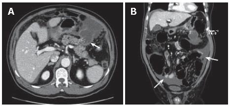
Figure 1 CT scan of case 1 showing a 5 cm-sized ruptured cystic mass in the pancreatic body and tail junction (arrow) (A) and massive ascites throughout the peritoneal space (arrow) (B).
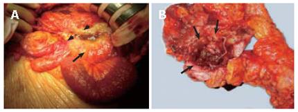
Figure 2 Operation of case 1 revealing a ruptured cystic mass at pancreatic tail (arrow) and spreaded mucinous materials around the mass (arrow heads) (A) and a ruptured mucinous cystic mass of pancreas (arrows) (B).
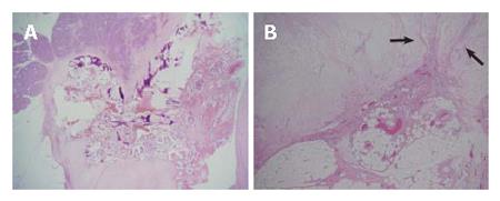
Figure 3 Microscopic examination of case 1 displaying papillary growth in the pancreatic duct (HE, × 12.
5) (A) and infiltration of malignant cells into the pancreas parenchyma (HE, × 40, arrows) (B).
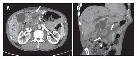
Figure 4 CT scan of case 2 demonstrating a 10 cm-sized cystic mass in pancreatic head (arrow) (A) and two communications between stomach and mass and between bulb and mass (arrows) and patulous ampulla of Vater (arrow head) (B).
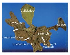
Figure 5 Specimen of case 2 showing two fistulous openings at stomach, duodenal bulb and patulous ampulla of Vater (arrows).
- Citation: Lee SE, Jang JY, Yang SH, Kim SW. Intraductal papillary mucinous carcinoma with atypical manifestations: Report of two cases. World J Gastroenterol 2007; 13(10): 1622-1625
- URL: https://www.wjgnet.com/1007-9327/full/v13/i10/1622.htm
- DOI: https://dx.doi.org/10.3748/wjg.v13.i10.1622


