Copyright
©2007 Baishideng Publishing Group Co.
World J Gastroenterol. Mar 14, 2007; 13(10): 1561-1568
Published online Mar 14, 2007. doi: 10.3748/wjg.v13.i10.1561
Published online Mar 14, 2007. doi: 10.3748/wjg.v13.i10.1561
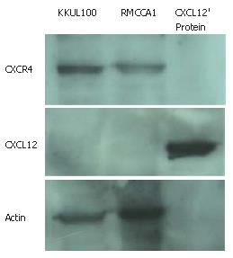
Figure 1 CXCR4 and CXCL12 expression in human colorectal carcinoma cell lines (RMCCA1 and KKU100).
Western Blot analysis revealed the presence of CXCR4 specific bands in both cell lines. However, no expression of CXCL12 was identified in both cell lines. 1The CXCL12 recombinant protein was used as a positive control for detection of CXCL12.
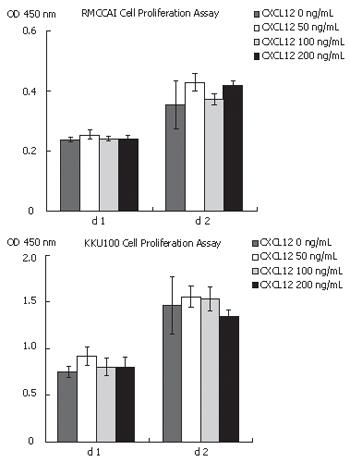
Figure 2 Effect of CXCL12 on the proliferation of cholangiocarcinoma cells.
RMCCA1 and KKU100 were treated with CXCL12 at various concentrations (0, 50, 100 and 200 ng/mL). Cell proliferation assay was performed after 2 d by using WST-1. The absorbance at 450 nm, against a reference wavelength of 650 nm, was determined.
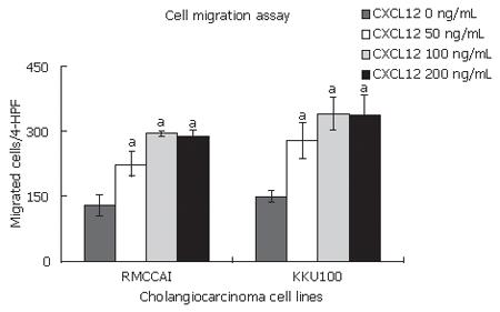
Figure 3 Effect of CXCL12 on the migration of cholangiocarcinoma cells.
RMCCA1 and KKU100 were seeded in the 8-μm pore filters (Transwell, 24-well cell culture, Coster, Boston, MA). The bottom chamber contained 0 or 100 ng/mL of CXCL12. After 24 h, the cells on the lower surface were counted under a microscope at five random 100 x power fields. The experiment was repeated for 3 times and the data represent the average results from 3 individual experiments. CXCL12 induced the migration of cholangiocarcinoma cells (aP < 0.05 vs 0 ng/mL of CXCL12).
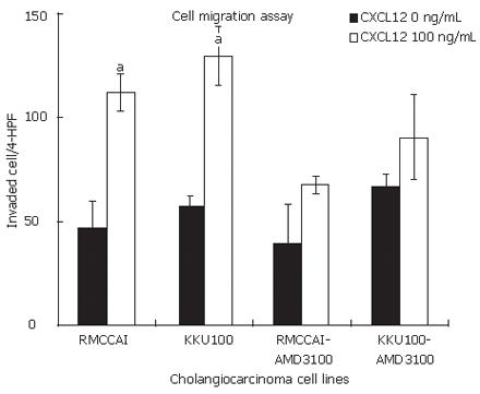
Figure 4 Effect of CXCL12 on the invasion of cholangiocarcinoma cells.
RMCCA1 and KKU100 were pre-treated with or without AMD3100 for 12 h then were seeded in the 24-well Biocoat Matrigel invasion chamber. The bottom chamber contained 0 or 100 ng/mL of CXCL12. After 24 h, the cells on the lower surface were assayed as described previously. CXCL12 induced the invasion of cholangiocarcinoma cells. The effect of CXCL12 was decreased when the cells were pre-treated with AMD3100 (aP < 0.05).
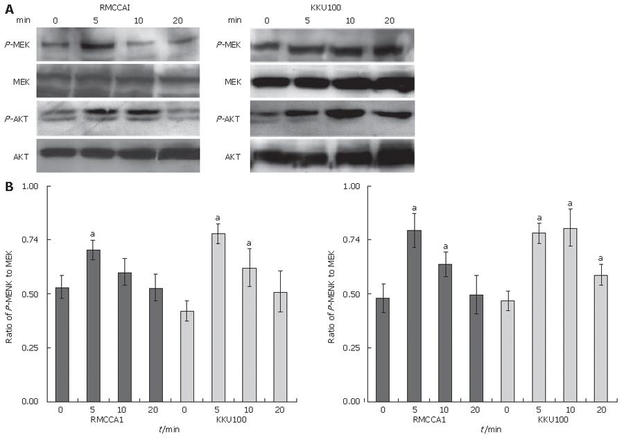
Figure 5 A: Western blot analysis of MEK1/2 and Akt phosphorylation in CXCL12-treated cholangiocarcinoma cells.
RMCCA1 and KKU 100 were treated with 100 ng/mL of CXCL12 for indicated time. MEK1/2 and Akt phosphorylation were determined by Western blot as described; B: The average band intensity based on 3 biologically separate experiments, showing levels of MEK1/2 and Akt phosphorylation relative to the levels of MEK1/2 and Akt expression from the same experiment (aP < 0.05).
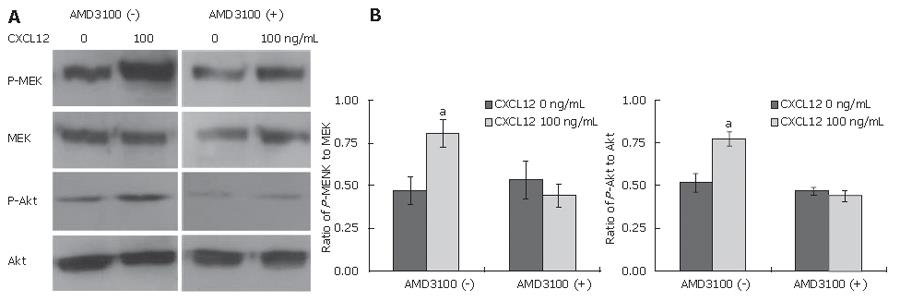
Figure 6 A: Effect of AMD3100 on CXCL12-induced phosphorylation of MEK1/2 and Akt in cholangiocarcinoma cells.
RMCCA1 cells were pre-treated with or without AMD3100 before added CXCL12. Cells were collected at 5 min and MEK1/2 and Akt phosphorylation were determined by Western blot as described; B: The average band intensity based on 3 biologically separate experiments, showing levels of MEK1/2 and Akt phosphorylation relative to the levels of MEK1/2 and Akt expression from the same experiment (aP < 0.05 vs 0 ng/mL of CXCL12).
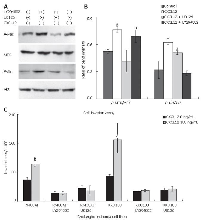
Figure 7 A: Effect of U0126 and LY294002 on CXCL12-induced phosphorylation of MEK1/2 and Akt in cholangiocarcinoma cells.
RMCCA1 cells were pre-treated with U0126 or LY294002 before added CXCL12. Cells were collected at 5 min and MEK1/2 and Akt phosphorylation were determined by Western blot as described; B: The average band intensity based on 3 biologically separate experiments, showing levels of MEK1/2 and Akt phosphorylation relative to the levels of MEK1/2 and Akt expression from the same experiment (aP < 0.05 compared with control); C: Effect of MEK1/2 and Akt phosphorylation induced by CXCL12 on the invasion of cholangiocarcinoma cells. RMCCA1 and KKU100 were pre-treated with or without LY294002 and U0126 for 12 h then were seeded in the 24-well Biocoat Matrigel invasion chamber. The bottom chamber contained 0 or 100 ng/mL of CXCL12. After 24 h, the cells on the lower surface were assayed as described previously. LY294002 and U0126 inhibited the effect of CXCL12 induced cholangiocarcinoma cells invasion (aP < 0.05).
Figure 8 Gelatin zymography of the conditioned medium from cholangiocarcinoma cell lines revealed the proteolytic bands at molecular weight indicating them to be MMP-9.
Levels of the proteolytic activity are not different between each sample.
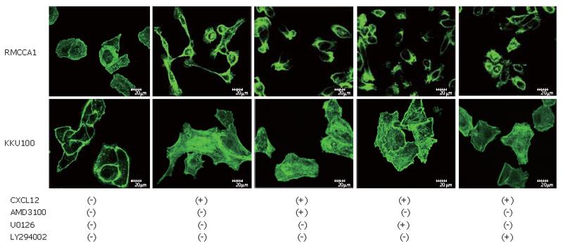
Figure 9 Effect of CXCL12 on the polymerization of actin cytoskeleton.
Cholangiocarcinoma cells were pre-treated with vehicle, AMD3100, U0126 or LY294002 and incubated for 6 h in medium containing 0 or 100 ng/mL CXCL12. The cells were stained with Alexa Fluor 488 Phalloidin to visualize actin cytoskeleton under a Confocal laser scanning microscope (Olympus SV1000).
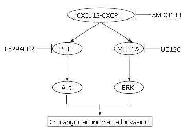
Figure 10 The pathway diagram identified the targets of AMD3100, U0126 and LY294002 (CXCR4, MEK1/2 and PI3K respectively).
Inhibition of these pathways abrogates the invasion of cholangiocarcinoma cells.
- Citation: Leelawat K, Leelawat S, Narong S, Hongeng S. Roles of the MEK1/2 and AKT pathways in CXCL12/CXCR4 induced cholangiocarcinoma cell invasion. World J Gastroenterol 2007; 13(10): 1561-1568
- URL: https://www.wjgnet.com/1007-9327/full/v13/i10/1561.htm
- DOI: https://dx.doi.org/10.3748/wjg.v13.i10.1561


