Copyright
©2007 Baishideng Publishing Group Co.
World J Gastroenterol. Jan 7, 2007; 13(1): 39-47
Published online Jan 7, 2007. doi: 10.3748/wjg.v13.i1.39
Published online Jan 7, 2007. doi: 10.3748/wjg.v13.i1.39
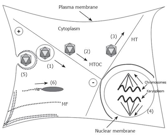
Figure 1 Participation of microtubules and microfilaments in transport processes.
Transport processes are indicated as bold arrows. Microtubules (MT, bold lines) have a highly dynamic plus-end and a less dynamic minus-end that is located at the microtubule-organizing (MTOC). They participate in the transport of (1) organelles, e.g. endosomes, (2) direct retrograde transport of capsids via the dynein motor protein complex (adenovirus, HSV 1, parvoviruses), (3) direct anterograde transport of progeny HSV 1 capsids by conventional kinesin, and (4) participate in chromosome segregation upon mitosis. Microfilaments (MF), depicted as dotted lines participate (5) in separation of endocytotic vesicles from the plasma membrane and (6) via polymerization in transport of e.g. Listeria monocytogenes and nuclear polyhedrosis virus (NPV).
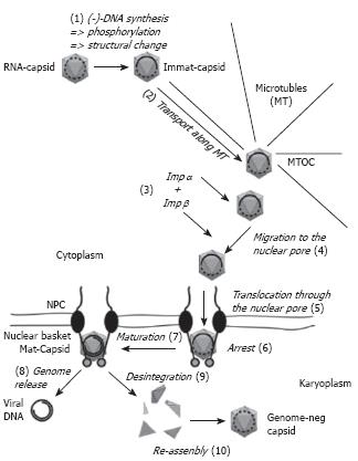
Figure 2 Hepadnaviral trafficking within the cell.
Capsids are drawn as grey icosahedra. Immat-Capsid, immature capsid, Mat-Capsid, mature capsid. The nucleic acid found within the capsids is depicted as a dotted line (RNA) or a full line (DNA). The arrows present movements (2, 4, 5, 8) or changes of the capsid (1, 7, 9, 10). Further explanations are given in the text.
- Citation: Kann M, Schmitz A, Rabe B. Intracellular transport of hepatitis B virus. World J Gastroenterol 2007; 13(1): 39-47
- URL: https://www.wjgnet.com/1007-9327/full/v13/i1/39.htm
- DOI: https://dx.doi.org/10.3748/wjg.v13.i1.39


