Copyright
©2006 Baishideng Publishing Group Co.
World J Gastroenterol. Aug 21, 2006; 12(31): 5051-5054
Published online Aug 21, 2006. doi: 10.3748/wjg.v12.i31.5051
Published online Aug 21, 2006. doi: 10.3748/wjg.v12.i31.5051
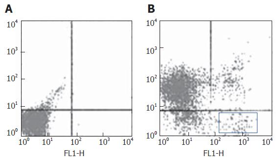
Figure 1 Bone marrow stem cells sorted by flow-cytometry.
A: Control; B: Percentage of Thy+CD3-CD45RA- cells in bone marrow cells without erythrocytes (about 2.8%).
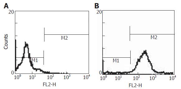
Figure 2 PKH26-GL labeling of Thy+CD3-CD45RA- cells.
A: Control; B: Percentage of PKH26-GL-labeled cells in Thy+CD3-CD45RA- cells. (M1: unlabeled cells; M2: labeled cells).
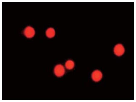
Figure 3 Red Thy+CD3-CD45RA- cells with PKH26-GL under fluorescence microscope.
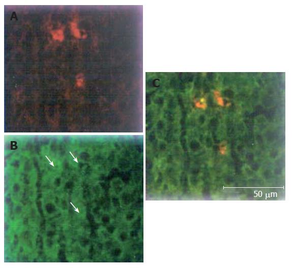
Figure 4 Red sporadic PKH26-GL-labeled cells (A), green hepatocytes (B), and yellow PKH26-GL-labeled cells expressing albumin (C).
- Citation: Zhan YT, Wang Y, Wei L, Liu B, Chen HS, Cong X, Fei R. Differentiation of rat bone marrow stem cells in liver after partial hepatectomy. World J Gastroenterol 2006; 12(31): 5051-5054
- URL: https://www.wjgnet.com/1007-9327/full/v12/i31/5051.htm
- DOI: https://dx.doi.org/10.3748/wjg.v12.i31.5051


