Copyright
©2006 Baishideng Publishing Group Co.
World J Gastroenterol. Jul 14, 2006; 12(26): 4181-4184
Published online Jul 14, 2006. doi: 10.3748/wjg.v12.i26.4181
Published online Jul 14, 2006. doi: 10.3748/wjg.v12.i26.4181
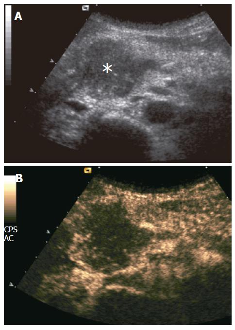
Figure 1 Ductal adenocarcinoma.
A: US showing slightly hypoechoic pancreatic head mass (asterisk); B: CEUS showing poor enhancement of the mass, appearing hypoechoic to the rest of pancreatic parenchyma in the contrast-enhanced phases.
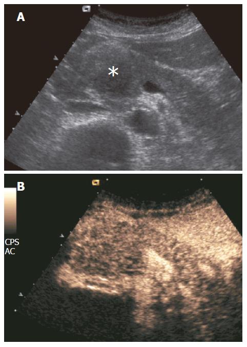
Figure 2 Mass-forming chronic autoimmune pancreatitis.
A: US showing hypoechoic head pancreatic mass (asterisk); B: CEUS showing parenchymographic enhancement of the pancreatic lesion in the head of the pancreas during the contrast-enhanced phases.
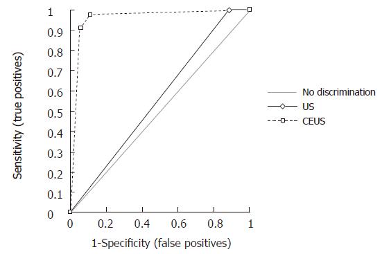
Figure 3 Receiver operating characteristic curves of baseline ultrasound and contrast-enhanced ultrasound in the characterization of 173 pancreatic masses, discerning between benignancy (pancreatitis) and malignancy (tumor).
- Citation: D’Onofrio M, Zamboni G, Tognolini A, Malagò R, Faccioli N, Frulloni L, Mucelli RP. Mass-forming pancreatitis: Value of contrast-enhanced ultrasonography. World J Gastroenterol 2006; 12(26): 4181-4184
- URL: https://www.wjgnet.com/1007-9327/full/v12/i26/4181.htm
- DOI: https://dx.doi.org/10.3748/wjg.v12.i26.4181


