Copyright
©2006 Baishideng Publishing Group Co.
World J Gastroenterol. Jun 7, 2006; 12(21): 3442-3445
Published online Jun 7, 2006. doi: 10.3748/wjg.v12.i21.3442
Published online Jun 7, 2006. doi: 10.3748/wjg.v12.i21.3442
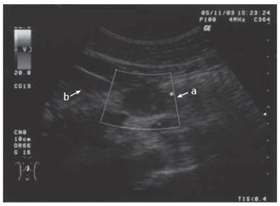
Figure 1 Echo tomography of gastric region.
A solid hypoechogenic mass with a diameter of 2.7 cm × 2.2 cm protruding from beyond the anterior wall of the stomach.
Figure 2 Macroscopic appearance of resected specimen: section of surgical specimen: a soft yellow mass of about 2 cm in diameter.
Figure 3 Histological findings of the resected specimen.
A: hematoxylin–eosin staining. Sheet of large and polygonal cells with round to oval, eccentrically located nuclei and eosinophilic granular cytoplasm with lower mitotic activity; B: immunohistochemical staining for S-100 protein. Diffuse and strong expression of S-100 protein; C: immunohistochemical staining for p53. Some of the tumor cells are positive for p53; D: immunohistochemical staining for Ki-67. Some of the tumor cells are positive for Ki-67.
- Citation: Patti R, Almasio PL, Vita GD. Granular cell tumor of stomach: A case report and review of literature. World J Gastroenterol 2006; 12(21): 3442-3445
- URL: https://www.wjgnet.com/1007-9327/full/v12/i21/3442.htm
- DOI: https://dx.doi.org/10.3748/wjg.v12.i21.3442


