Copyright
©2006 Baishideng Publishing Group Co.
World J Gastroenterol. Jun 7, 2006; 12(21): 3368-3372
Published online Jun 7, 2006. doi: 10.3748/wjg.v12.i21.3368
Published online Jun 7, 2006. doi: 10.3748/wjg.v12.i21.3368
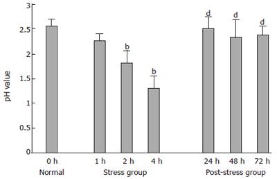
Figure 1 pH value of gastric juice in all groups.
bP < 0.01 vs normal group; dP < 0.01 vs stress 4 h group.
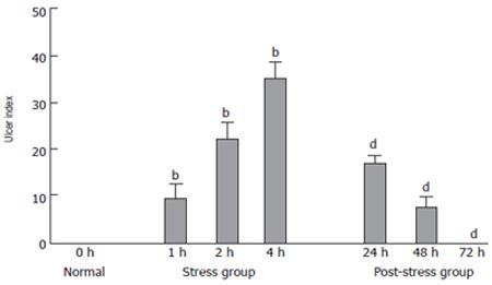
Figure 2 Ulcer index (UI) of gastric mucosa in all groups.
bP < 0.01 vs normal group; dP < 0.01 vs stress 4 h group.
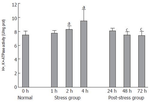
Figure 3 H+, K+-ATPase activity of parietal cells in all groups.
aP < 0.05 vs normal group; cP < 0.05 vs stress 4 h group.
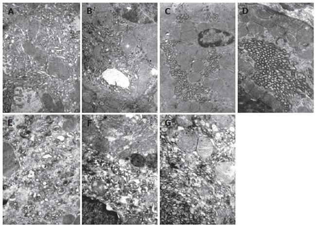
Figure 4 Ultrastructural change of parietal cells.
A: Gastric parietal cells presented resting state, abundant tubulovesicles and few intracellular canaliculi lined with rare microvilli could be found in the cytoplasm in normal group (×12 000); B: Parietal cells presented pre-secreting state, intracellular canaliculi lined with short microvilli were observed, plenty of vesicles were located around the canaliculi and accumulated on the apical plasma membrane in 1 h WRS group (×12 000); C: Parietal cells presented secreting state with decreased vesicles and increased intracellular canaliculi lined with elongated microvilli in 2 h WRS group; D: Parietal cells presented active secreting state and cytoplasm was almost devoid of vesicles and intracellular canalicular lumen contained numerous elongated microvilli in 4 h WRS group (×12 000); E - G: at 24, 48 and 72 h at the end of WRS for 4 h, Parietal cells returned to resting state with abundant tubulovesicles and few canaliculi in cytoplasm in post-stress group swollen mitochondria, vacuolar degeneration, crista-fragmentation, concentrated karyotin, roughness of nuclear envelopes and dilated perinuclear space were observed ( ×15 000).
- Citation: Li YM, Lu GM, Zou XP, Li ZS, Peng GY, Fang DC. Dynamic functional and ultrastructural changes of gastric parietal cells induced by water immersion-restraint stress in rats. World J Gastroenterol 2006; 12(21): 3368-3372
- URL: https://www.wjgnet.com/1007-9327/full/v12/i21/3368.htm
- DOI: https://dx.doi.org/10.3748/wjg.v12.i21.3368


