Copyright
©2006 Baishideng Publishing Group Co.
World J Gastroenterol. Jun 7, 2006; 12(21): 3324-3331
Published online Jun 7, 2006. doi: 10.3748/wjg.v12.i21.3324
Published online Jun 7, 2006. doi: 10.3748/wjg.v12.i21.3324
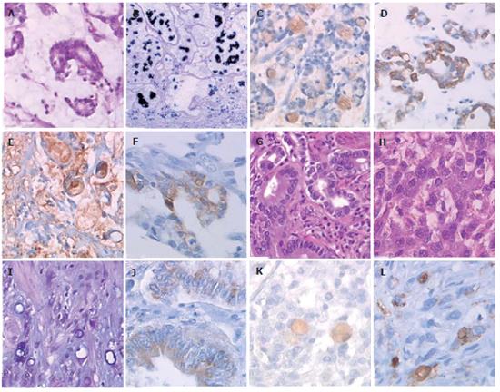
Figure 1 Expression of the secreted gel-forming mucins in gastric cancers.
A: Mucinous subtype in WHO classification, group II in Goseki’s classification (HE-safron × 125); B: Strong MUC2 in mucinous subtype ( in situ hybridization × 40); C: MUC2 in intracellular mucus vacuoles in mucinous subtype (SABC × 125); D: MUC5AC in the cytoplasm of tumoral cells in mucinous subtype (SABC × 125); E: MUC5B in the cytoplasm, within intracellular mucus vacuoles and in extracellular mucus in mucinous subtype (SABC × 125); F: MUC6 in the cytoplasm of tumoral cells in mucinous subtype (SABC × 200); G: Gastric cancer classified group I in Goseki’s classification (HE-safron × 125); H: Gastric cancer classified group III in Goseki’s classification (HE-safron, × 200); I: Gastric cancer classified group IV in Goseki’s classification (PAS and Alcian blue × 200); J: MUC2 in the cytoplasm of tumoral cells in group I gastric cancer Goseki’s classification (SABC × 200); K: MUC2 in intracellular mucus vacuoles in group III gastric cancer in Goseki’s classification (SABC × 200); L: MUC2 in intracellular mucus vacuoles in group IV gastric cancer in Goseki’s classification (SABC × 200).
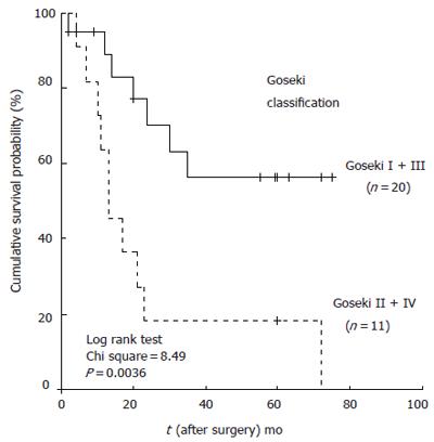
Figure 2 Survival curves for patients divided according to their Goseki classification.
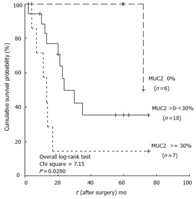
Figure 3 Survival curves for patients divided according to their immunohistochemical MUC2 expression.
- Citation: Leteurtre E, Zerimech F, Piessen G, Wacrenier A, Leroy X, Copin MC, Mariette C, Aubert JP, Porchet N, Buisine MP. Relationships between mucinous gastric carcinoma, MUC2 expression and survival. World J Gastroenterol 2006; 12(21): 3324-3331
- URL: https://www.wjgnet.com/1007-9327/full/v12/i21/3324.htm
- DOI: https://dx.doi.org/10.3748/wjg.v12.i21.3324


