Copyright
©2006 Baishideng Publishing Group Co.
World J Gastroenterol. Mar 21, 2006; 12(11): 1798-1801
Published online Mar 21, 2006. doi: 10.3748/wjg.v12.i11.1798
Published online Mar 21, 2006. doi: 10.3748/wjg.v12.i11.1798
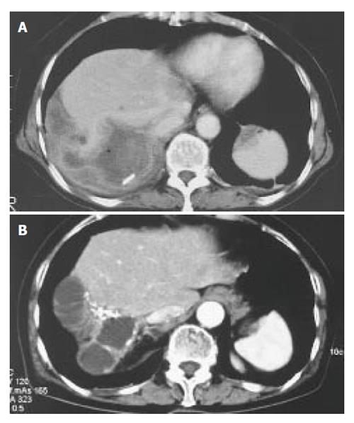
Figure 1 Computed tomography revealed a huge multilocular cyst measuring 10 cm in the right lobe of the liver on September 11th, 1997 (A).
A similar cyst with a calcified lesion was found in the right lobe of the liver on May 15th, 2003 (B).
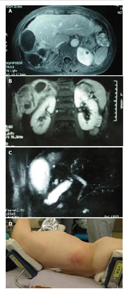
Figure 2 An axial (A) and coronal (B) view of magnetic resonance imaging (MRI) showed a multilocular cyst with a thick wall with a solid component extending into the subcutaneous tissue.
MR cholangio-pancreatography showed no dilatation in the biliary system, and most likely no communication with the liver cyst (C). The skin showed red swelling by the subcutaneous extension of the hepatic cyst (D).
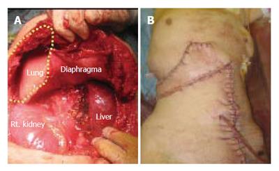
Figure 3 An en bloc resection resulted in a huge defect (A, yellow dotted circle).
The defect of the diaphragm was covered with the great omentum, and the thoraco abdominal wall defect was reconstructed by a musculo-cutaneous flap including the anterior sheath and the left rectus abdominis muscle which both receive the blood supply comes from the superior abdominal artery (B).
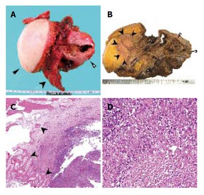
Figure 4 Macroscopically, the cystic mass directly invaded the intercostal space through the diaphragm (A.
Resected crude specimen, black triangle; skin, black arrow head; ribs, white triangle; cystic tumor; B. Cut surface of the specimen, black arrow head; subcutaneous invasion, white triangle; cystic tumor). Microscopically, the bone was destroyed by an invasion of granuloma tissue (C). The granuloma was composed of epithelioid cells with necrotic areas (D).
- Citation: Kawashita Y, Kamohara Y, Furui J, Fujita F, Miyamoto S, Takatsuki M, Abe K, Hayashi T, Ohno Y, Kanematsu T. Destructive granuloma derived from a liver cyst: A case report. World J Gastroenterol 2006; 12(11): 1798-1801
- URL: https://www.wjgnet.com/1007-9327/full/v12/i11/1798.htm
- DOI: https://dx.doi.org/10.3748/wjg.v12.i11.1798


