Copyright
©2006 Baishideng Publishing Group Co.
World J Gastroenterol. Mar 21, 2006; 12(11): 1774-1779
Published online Mar 21, 2006. doi: 10.3748/wjg.v12.i11.1774
Published online Mar 21, 2006. doi: 10.3748/wjg.v12.i11.1774
Figure 1 Expression of mda-7/IL-24 mRNA.
Infection of normal human liver cells and HCC with Ad.mda-7 resulted in an expression of mda-7/IL-24 mRNA. Cells infected with 1 000 VP/cell of Ad.vec or Ad.mda-7 were harvested at 48 h, treated as described in “Materials and Methods”. The total RNA was extracted and the RT-PCR was performed. (A) Lane 1: marker; lane 2: control L02 cells; lane 3: Ad.vec-infected L02 cells; lane 4: Ad.mda-7-infected L02 cells; lane 5: control HepG2 cells; lane 6: Ad.vec-infected HepG2 cells; lane 7: Ad.mda-7-infected HepG2 cells. (B) Expression of β-actin.
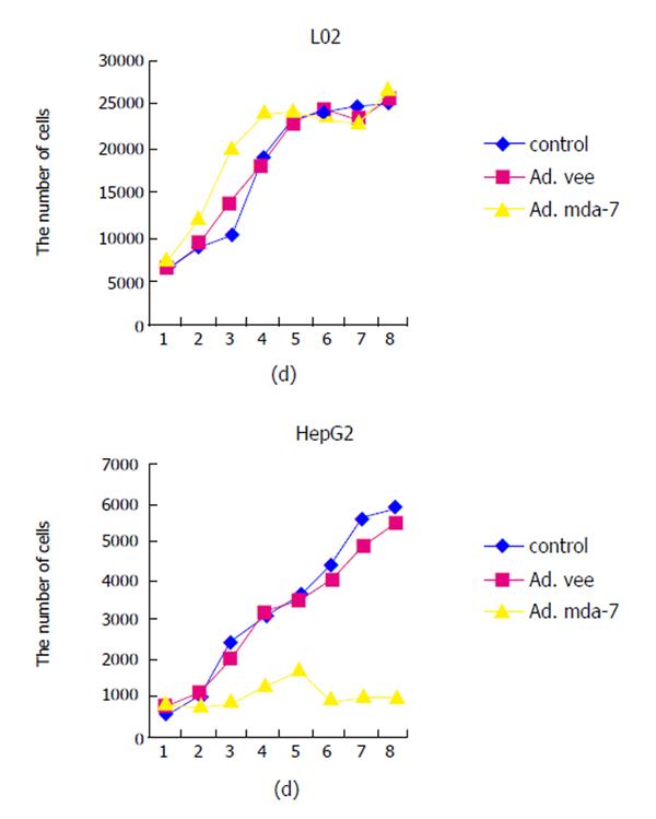
Figure 2 Ad.
mda-7 expression causing in vitro inhibition of growth of HCC cells, but not normal liver cells. The various cell types were uninfected (control) or infected with 1 000 VP/cell of Ad.vec or Ad.mda-7. Cell numbers were determined over an 8-d period. Cells were seeded in 96-well plates and treated the next day as described in ‘Materials and Methods’. After 24 h, the medium was removed, and cells were stained with MTT. Experiments were repeated three times in quadruplicates, and the results were presented as mean±SE.
Figure 3 Over-expression of mda-7 causes apoptosis selectively in HCC cells (400×).
Cells were infected with 1 000 VP/cell of Ad.mda-7 and all assays were performed 48 h after infection as described in ‘Materials and Methods’. To evaluate characteristic apoptotic morphology with fluorescence microscopy, the cells were seeded on glass slides and fixed with 40 g/L paraformaldehyde in PBS for 1 h at room temperature. After washing twice with PBS, cells were stained for 30 min in the dark at room temperature with 0.05 mg/mL Hoechst 33258 in PBS. The nuclear fragmentations were visualized by using a fluorescence microscope equipped with a UV-2A filter and Olympus BX60 photographic camera. Apoptotic cells were recognized by condensation of nuclear chromatin and its fragmentation.
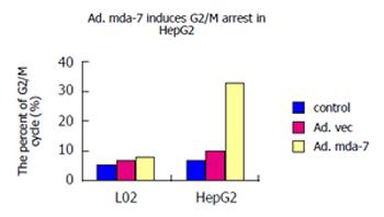
Figure 4 Induction of G2/M arrest in HCC cells HepG2, but not in normal liver cells L02 by Ad.
mda-7 infection.
- Citation: Wang CJ, Xue XB, Yi JL, Chen K, Zheng JW, Wang J, Zeng JP, Xu RH. Melanoma differentiation-associated gene-7, MDA-7/IL-24, selectively induces growth suppression, apoptosis in human hepatocellular carcinoma cell line HepG2 by replication-incompetent adenovirus vector. World J Gastroenterol 2006; 12(11): 1774-1779
- URL: https://www.wjgnet.com/1007-9327/full/v12/i11/1774.htm
- DOI: https://dx.doi.org/10.3748/wjg.v12.i11.1774


