Copyright
©2006 Baishideng Publishing Group Co.
World J Gastroenterol. Mar 21, 2006; 12(11): 1757-1760
Published online Mar 21, 2006. doi: 10.3748/wjg.v12.i11.1757
Published online Mar 21, 2006. doi: 10.3748/wjg.v12.i11.1757
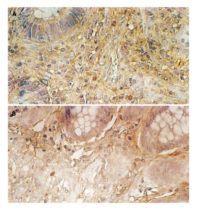
Figure 1 TIDCs in earlier (A) and later (B) stages of rectal cancer.
(magnification: A, B x 400)
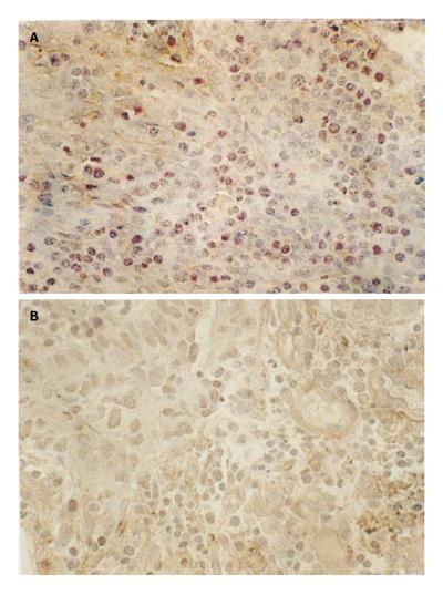
Figure 2 TILs in earlier (A) and later (B) stages of rectal cancer.
(magnification: A, B x 400)
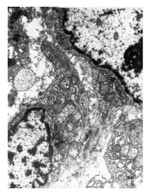
Figure 3 Morphology of TIDCs .
D represents TIDCs, T represents tumor cells. (magnification x 10000)
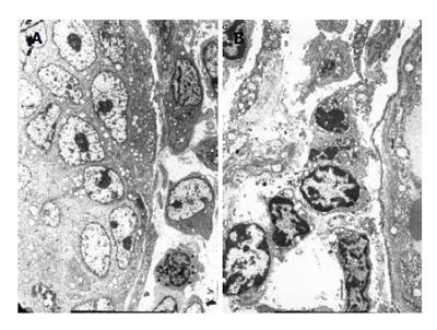
Figure 4 Lymphocytes in earlier (A) and later (B) stages of rectal cancer.
(magnification: A x4000, B x2000)
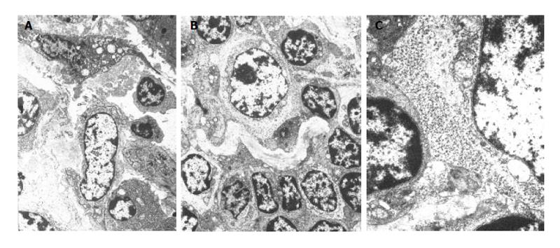
Figure 5 Relations between TILs and tumors cells (A), TIDCs and TILs (B) and glycogen granules (C).
(magnification: A, x 4000, B x3500, C x15000)
- Citation: Xie ZJ, Jia LM, He YC, Gao JT. Morphological observation of tumor infiltrating immunocytes in human rectal cancer. World J Gastroenterol 2006; 12(11): 1757-1760
- URL: https://www.wjgnet.com/1007-9327/full/v12/i11/1757.htm
- DOI: https://dx.doi.org/10.3748/wjg.v12.i11.1757


