Copyright
©2005 Baishideng Publishing Group Inc.
World J Gastroenterol. Nov 28, 2005; 11(44): 7048-7050
Published online Nov 28, 2005. doi: 10.3748/wjg.v11.i44.7048
Published online Nov 28, 2005. doi: 10.3748/wjg.v11.i44.7048
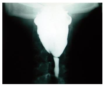
Figure 1 Esophagography showing a linear filling defect (1.
5 cm length) at the upper esophagus, suggesting esophageal web.
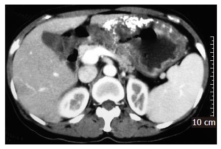
Figure 2 Contrast-enhanced abdominal CT scan showing diffuse irregular wall thickness with multiple calcifications at the body and antrum of the stomach.
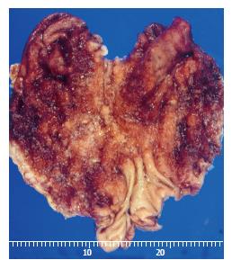
Figure 3 Macroscopic appearance of resected stomach showing a diffuse large Borrmann type 4 cancer in the whole body and cardia.
- Citation: Kim KH, Kim MC, Jung GJ. Gastric cancer occurring in a patient with Plummer-Vinson syndrome: A case report. World J Gastroenterol 2005; 11(44): 7048-7050
- URL: https://www.wjgnet.com/1007-9327/full/v11/i44/7048.htm
- DOI: https://dx.doi.org/10.3748/wjg.v11.i44.7048


