Copyright
©2005 Baishideng Publishing Group Inc.
World J Gastroenterol. Nov 7, 2005; 11(41): 6557-6559
Published online Nov 7, 2005. doi: 10.3748/wjg.v11.i41.6557
Published online Nov 7, 2005. doi: 10.3748/wjg.v11.i41.6557
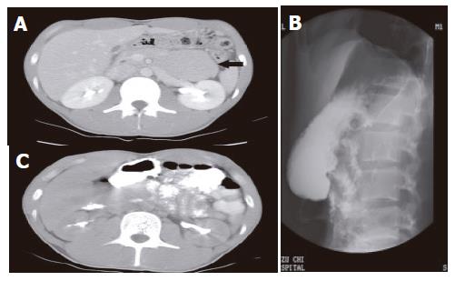
Figure 1 A: Computed tomography (CT) showing a soft tissue mass between the stomach, the pancreas, and the transverse colon (arrow); B: Upper gastrointestinal series showing a cluster of jejunum interposing between the stomach and the spine; C: The mass proved to be a cluster of intestinal loop after ingestion of contrast medium.
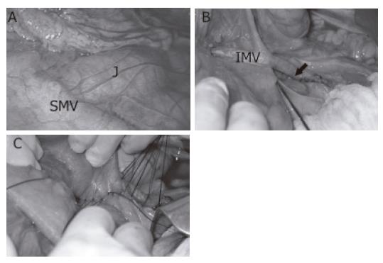
Figure 2 A: Loops of small bowel behind the mesocolon.
Treitz’s ligament was not seen. SMV: superior mesenteric vein, J: jejunum; B: the orifice through which the small bowel herniated (arrow). IMV: inferior mesenteric vein; C: completion of the repair.
- Citation: Huang YM, Chou ASB, Wu YK, Wu CC, Lee MC, Chen HT, Chang YJ. Left paraduodenal hernia presenting as recurrent small bowel obstruction. World J Gastroenterol 2005; 11(41): 6557-6559
- URL: https://www.wjgnet.com/1007-9327/full/v11/i41/6557.htm
- DOI: https://dx.doi.org/10.3748/wjg.v11.i41.6557


