Copyright
©The Author(s) 2005.
World J Gastroenterol. Oct 14, 2005; 11(38): 6014-6017
Published online Oct 14, 2005. doi: 10.3748/wjg.v11.i38.6014
Published online Oct 14, 2005. doi: 10.3748/wjg.v11.i38.6014
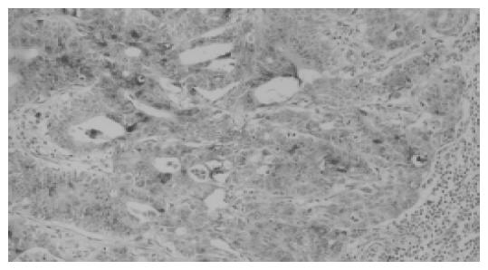
Figure 1 Typical result of immunohistochemical staining of RCAS1 (200) in metastatic lymph nodes.
Expression of RCAS1 was demonstrated both in cytoplasm and on cell membrane of tumor cells.
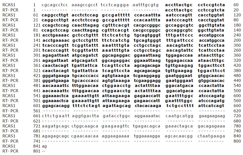
Figure 2 Nucleotide sequence alignment of RCAS1 cDNA from databases present at NCBI GenBank (gi: 13528905) and RT-PCR products from metastatic lymph nodes.
The residues boxed in black indicate positional identity for both sequences. Numbers of nucleotide sequences are indicated on the left and right margins.
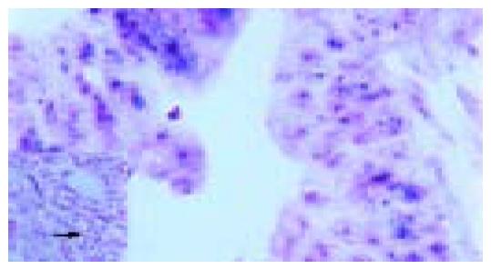
Figure 3 RCAS1 mRNA in situ hybridization in metastatic lymph nodes from colon cancer.
The positive signal was identified in the cytoplasm of tumor cells. All leukocytes showed negative signal for RCAS1 (as presented in the Figure).
- Citation: Leelawat K, Engprasert S, Tujinda S, Suthippintawong C, Enjoji M, Nakashima M, Watanabe T, Leardkamolkarn V. Receptor-binding cancer antigen expressed on SiSo cells can be detected in metastatic lymph nodes from gastrointestinal cancers. World J Gastroenterol 2005; 11(38): 6014-6017
- URL: https://www.wjgnet.com/1007-9327/full/v11/i38/6014.htm
- DOI: https://dx.doi.org/10.3748/wjg.v11.i38.6014


