Copyright
©The Author(s) 2005.
World J Gastroenterol. Aug 28, 2005; 11(32): 5068-5071
Published online Aug 28, 2005. doi: 10.3748/wjg.v11.i32.5068
Published online Aug 28, 2005. doi: 10.3748/wjg.v11.i32.5068
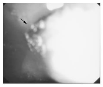
Figure 1 Endoscopic image: deformed duodenal bulb with slit-like opening (black arrow).
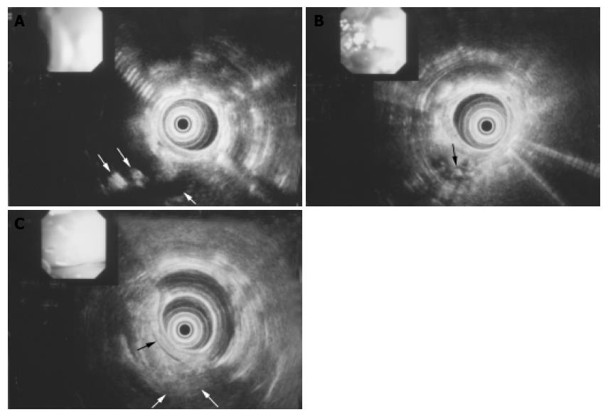
Figure 2 EUS image.
A: EUS image: dilated common bile duct (thick arrow) with stones and air (thin arrows) draining in the bulb; B: EUS image: dilated common bile duct with stones (black arrow) in the lumen; C: EUS image: unchanged main pancreatic duct (black arrow) with parenchymal irregularity (white arrows).
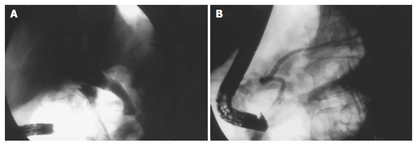
Figure 3 ERCP image.
A: ERCP image: dilated biliary tree filled with stones cannulated from the duodenal bulb; B: ERCP image: the main pancreatic duct making the loop in the pancreatic head.
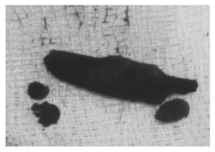
Figure 4 Extracted biliary stones.
- Citation: Krstic M, Stimec B, Krstic R, Ugljesic M, Knezevic S, Jovanovic I. EUS diagnosis of ectopic opening of the common bile duct in the duodenal bulb: A case report. World J Gastroenterol 2005; 11(32): 5068-5071
- URL: https://www.wjgnet.com/1007-9327/full/v11/i32/5068.htm
- DOI: https://dx.doi.org/10.3748/wjg.v11.i32.5068


