Copyright
©The Author(s) 2005.
World J Gastroenterol. Aug 28, 2005; 11(32): 4986-4991
Published online Aug 28, 2005. doi: 10.3748/wjg.v11.i32.4986
Published online Aug 28, 2005. doi: 10.3748/wjg.v11.i32.4986
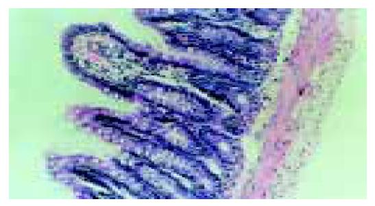
Figure 1 Normal villus and glands in normal group.
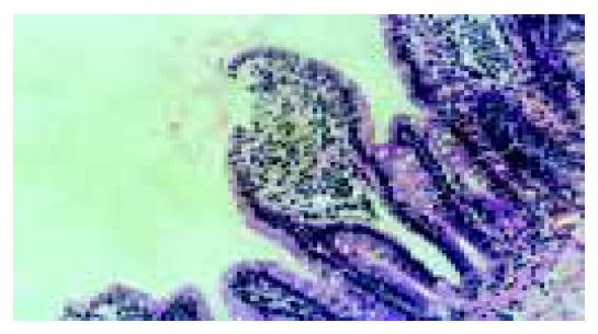
Figure 2 Severe edema of mucosa villus and infiltration of necrotic epithelial cells and inflammatory cells in model group.
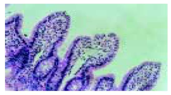
Figure 3 Light edema of mucosa villus and few necrotic epithelial cells and infiltration of few inflammatory cells in low dose group.
Figure 4 No significant edema and necrotic mucosa villus and infiltration of a few inflammatory cells in high dose group.
-
Citation: Hei ZQ, Huang HQ, Zhang JJ, Chen BX, Li XY. Protective effect of
Astragalus membranaceus on intestinal mucosa reperfusion injury after hemorrhagic shock in rats. World J Gastroenterol 2005; 11(32): 4986-4991 - URL: https://www.wjgnet.com/1007-9327/full/v11/i32/4986.htm
- DOI: https://dx.doi.org/10.3748/wjg.v11.i32.4986


