Copyright
©2005 Baishideng Publishing Group Co.
World J Gastroenterol. Jan 21, 2005; 11(3): 348-352
Published online Jan 21, 2005. doi: 10.3748/wjg.v11.i3.348
Published online Jan 21, 2005. doi: 10.3748/wjg.v11.i3.348
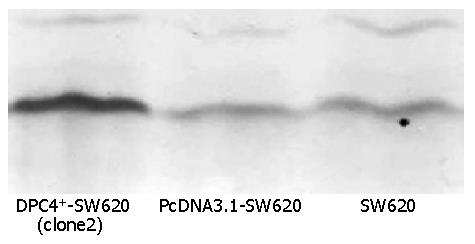
Figure 1 Strongest expression of Smad4 protein in clone2 detected by Western blot analysis.
Figure 2 Expression of smad4 protein.
A: Weak intracellular expression of Smad4 protein in SW620 cells. The positive staining for Smad4 was localized in cytoplasm; B: Weak intracellular expression of Smad4 protein in PcDNA3.1-SW620 cells. The positive staining for Smad4 was localized in cytoplasm; C: Strong intracellular expression of Smad4 protein in DPC4+-SW620 cells (clone2). The positive staining for Smad4 was localized in cytoplasm and nucleus, mainly in cytoplasm.
Figure 3 Growth property of different groups.
A: Growth curve of cells of different groups; B: Doubling time of cells of different groups; C: Cloning efficiencies in different groups. bP<0.01 vs DPC4+-SW620.
Figure 4 Alterations of S% (A) and apoptosis rate (B) detected by flow- cytometry.
aP<0.05 vs DPC4+-SW620.
- Citation: Xiao DS, Wen JF, Li JH, Hu ZL, Zheng H, Fu CY. Effect of deleted pancreatic cancer locus 4 gene transfection on biological behaviors of human colorectal carcinoma cells. World J Gastroenterol 2005; 11(3): 348-352
- URL: https://www.wjgnet.com/1007-9327/full/v11/i3/348.htm
- DOI: https://dx.doi.org/10.3748/wjg.v11.i3.348


