Copyright
©The Author(s) 2005.
World J Gastroenterol. Jul 28, 2005; 11(28): 4443-4444
Published online Jul 28, 2005. doi: 10.3748/wjg.v11.i28.4443
Published online Jul 28, 2005. doi: 10.3748/wjg.v11.i28.4443
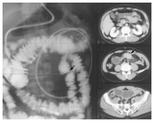
Figure 1 Small bowel series and CT showed both the distention of fluid- and gas-filled loops of small intestine and segmental stenosis indicated by arrow.
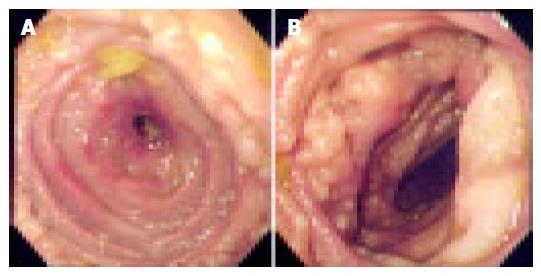
Figure 2 Resected jejunum show.
A: Resected jejunum showed diffuse thickening of the intestinal wall (oral side); B: multiple hyperplastic follicles (anal side).
- Citation: Nomura K, Tomikashi K, Matsumoto Y, Yoshida N, Okuda T, Sakakura C, Mitsufuji S, Horiike S, Yamagishi H, Okanoue T, Taniwaki M. Small bowel non-Hodgkin's lymphoma remaining in complete remission by surgical resection and adjuvant rituximab therapy. World J Gastroenterol 2005; 11(28): 4443-4444
- URL: https://www.wjgnet.com/1007-9327/full/v11/i28/4443.htm
- DOI: https://dx.doi.org/10.3748/wjg.v11.i28.4443


