Copyright
©The Author(s) 2005.
World J Gastroenterol. Jul 14, 2005; 11(26): 3985-3989
Published online Jul 14, 2005. doi: 10.3748/wjg.v11.i26.3985
Published online Jul 14, 2005. doi: 10.3748/wjg.v11.i26.3985
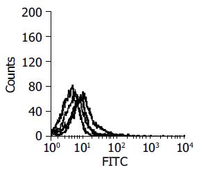
Figure 1 Result of polyclonal scFv phages by FCM (from left to right: purple -negative; green-1st round; red-2nd round; blue-3rd round).
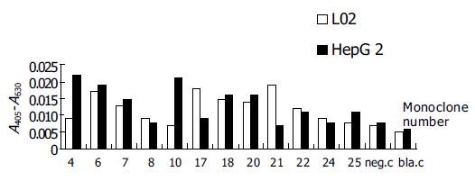
Figure 2 ELISA for monoclonal scFv phages.
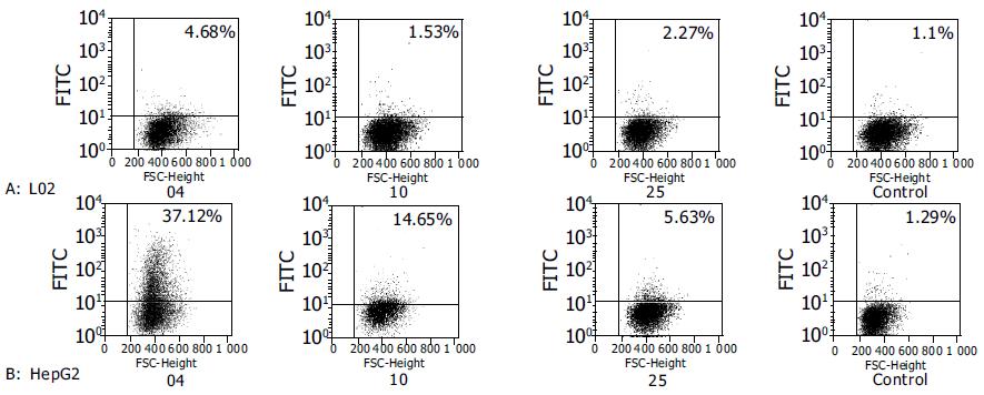
Figure 3 FCM for monoclonal scFv phages.
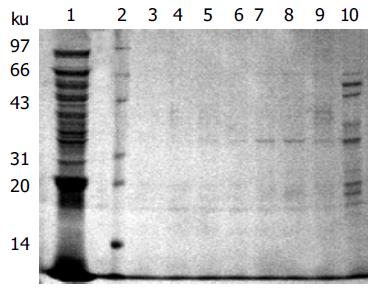
Figure 4 SLH04 scFv antibody fragments eluted with different concentrations of imidazole.
Lane 1: periplasm before eluting; lane 2: protein marker; lane 3, 4, 5, 6, 7, 8, 9, 10: elution washings at different imidazole concentrations, 200, 150, 100, 80, 60, 40, 20, 10 mmol/L, respectively.
Figure 5 Western blot of purified scFv antibodies SLH10 and SLH04.
Lane 1: protein marker; lane 2: purified scFv antibody SLH10; lane 3: purified scFv antibody SLH04; lane 4: the induced bacterial periplasmic of HB2151; lane 5: induced bacterial periplasm of HB2151 with pHEN2 vector.
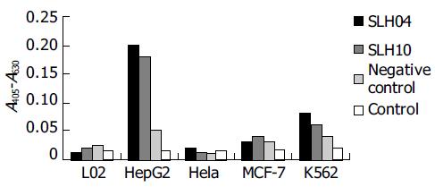
Figure 6 Two purified soluble scFv antibody fragments cells ELISA.
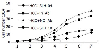
Figure 7 Proliferation of HepG2 cell inhibited by purified antibody fragments.
- Citation: Yu B, Ni M, Li WH, Lei P, Xing W, Xiao DW, Huang Y, Tang ZJ, Zhu HF, Shen GX. Human scFv antibody fragments specific for hepatocellular carcinoma selected from a phage display library. World J Gastroenterol 2005; 11(26): 3985-3989
- URL: https://www.wjgnet.com/1007-9327/full/v11/i26/3985.htm
- DOI: https://dx.doi.org/10.3748/wjg.v11.i26.3985


