Copyright
©2005 Baishideng Publishing Group Inc.
World J Gastroenterol. May 28, 2005; 11(20): 3167-3169
Published online May 28, 2005. doi: 10.3748/wjg.v11.i20.3167
Published online May 28, 2005. doi: 10.3748/wjg.v11.i20.3167
Figure 1 A: The preoperative examination: Colonoscopy showed a yellowish hemispherical tumor at the site of ascending colon.
The overlying mucosa was smooth, and the lesion was soft and compressible; B: The preoperative examination: Barium enema revealed an ovoid filling defect with smooth border at the proximal area of colon hepatic flexture; C: The preoperative examination: CT scan found a pedunculated neoplasm protruding into the lumen at the site of ascending colon with sharp margin and soft tissue density.
Figure 2 A: The macroscopic inspection: Macroscopic inspection of the resected colon segment showed a smooth round polypoid submucous tumor with elastic character and 35 mm×30 mm×24 mm in size; B: The macroscopic inspection: Fault of the specimen demonstrated the tumor with pedunculated appearance.
The base of the lesion was 8 mm in size. The tumor was covered by normal mucosa and had uniform parenchyma in bright yellow color (one scale mark = 1 mm).
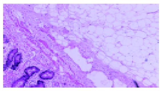
Figure 3 The histological examination: Histological examination showed characteristic lipoma of colon.
No evidence of malignancy was detected (hematoxylin and eosin, original magnification ×40).
- Citation: Zhang H, Cong JC, Chen CS, Qiao L, Liu EQ. Submucous colon lipoma: A case report and review of the literature. World J Gastroenterol 2005; 11(20): 3167-3169
- URL: https://www.wjgnet.com/1007-9327/full/v11/i20/3167.htm
- DOI: https://dx.doi.org/10.3748/wjg.v11.i20.3167


