Copyright
©2005 Baishideng Publishing Group Inc.
World J Gastroenterol. May 28, 2005; 11(20): 3161-3164
Published online May 28, 2005. doi: 10.3748/wjg.v11.i20.3161
Published online May 28, 2005. doi: 10.3748/wjg.v11.i20.3161
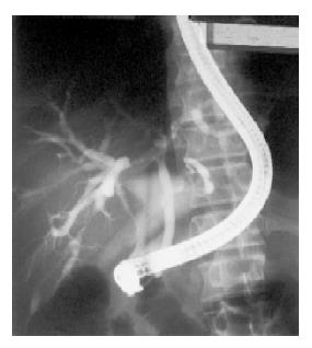
Figure 1 Endoscopic retrograde cholangiopancreatography showing bile duct stricture at liver hilum suggesting hilar cholangiocarcinoma.
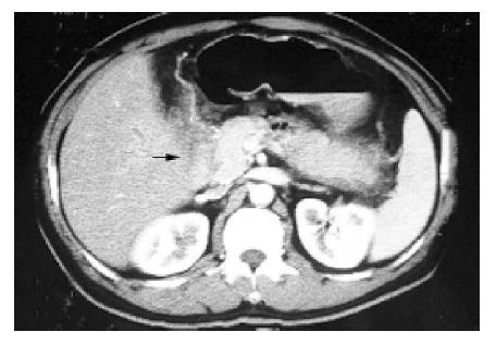
Figure 2 Contrast CT scan showing lymph node enlargement at hepatoduodenal ligament (black arrow).
Figure 3 Metallic stent (black arrow) inside common duct on endoscopic retrograde cholangiopancreatography.
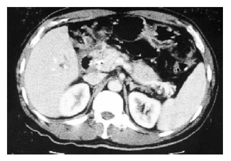
Figure 4 Contrast CT scan showing tumor regression after combined ILBT and external beam irradiation.
- Citation: Chan SY, Poon RT, Ng KK, Liu CL, Chan RT, Fan ST. Long-term survival after intraluminal brachytherapy for inoperable hilar cholangiocarcinoma: A case report. World J Gastroenterol 2005; 11(20): 3161-3164
- URL: https://www.wjgnet.com/1007-9327/full/v11/i20/3161.htm
- DOI: https://dx.doi.org/10.3748/wjg.v11.i20.3161


