Copyright
©2005 Baishideng Publishing Group Inc.
World J Gastroenterol. May 28, 2005; 11(20): 3151-3155
Published online May 28, 2005. doi: 10.3748/wjg.v11.i20.3151
Published online May 28, 2005. doi: 10.3748/wjg.v11.i20.3151
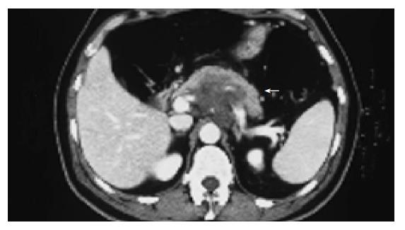
Figure 1 Basal contrast-enhanced abdominal CT scan.
The large retroperitoneal mass is indicated by the white arrow.
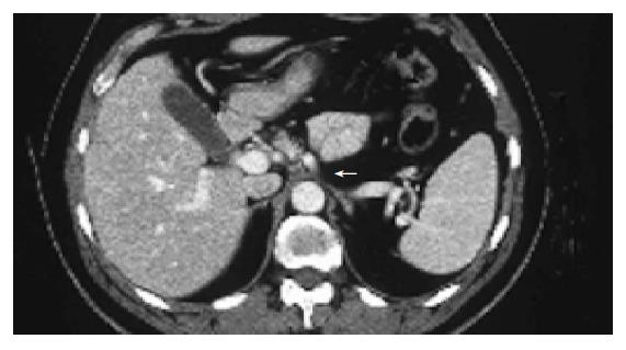
Figure 2 Contrast-enhanced abdominal CT scan performed.
The large retroperitoneal mass is reduced in size but a residual disease of about 1 cm is localized at celiac trunk (white arrow).
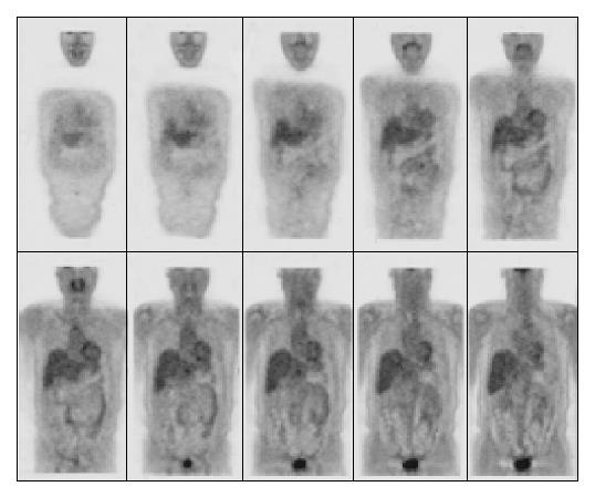
Figure 3 Total-body FDG-PET performed after the seventh course of treatment.
Figure 4 Contrast-enhanced CT performed 4 mo after the completion of the treatment.
A minimal residual disease is localized at the celiac tripod.
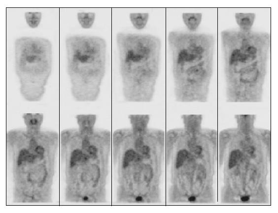
Figure 5 FDG-PET performed in October 2003: a complete remission of the disease is showed.
- Citation: Fulignati C, Pantaleo P, Cipriani G, Turrini M, Nicastro R, Mazzanti R, Neri B. An uncommon clinical presentation of retroperitoneal non-Hodgkin lymphoma successfully treated with chemotherapy: A case report. World J Gastroenterol 2005; 11(20): 3151-3155
- URL: https://www.wjgnet.com/1007-9327/full/v11/i20/3151.htm
- DOI: https://dx.doi.org/10.3748/wjg.v11.i20.3151


