Copyright
©2005 Baishideng Publishing Group Inc.
World J Gastroenterol. May 28, 2005; 11(20): 3046-3050
Published online May 28, 2005. doi: 10.3748/wjg.v11.i20.3046
Published online May 28, 2005. doi: 10.3748/wjg.v11.i20.3046
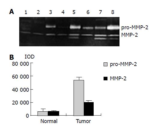
Figure 1 MMP-2 and pro-MMP-2 activity in normal and colorectal carcinoma tissues.
A: Results of gelatin zymography. Lanes 1-4: normal tissues; lanes 5-8: colorectal carcinoma tissues; B: Densitometric intensity of absorbance (IOD) of pro-MMP-2 and MMP-2 activity in normal and colorectal carcinoma tissues.
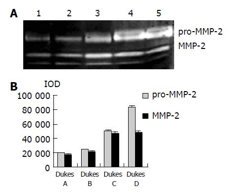
Figure 2 MMP-2 and pro-MMP-2 activity in colorectal carcinoma tissues at different Duke’s stage.
A: Results of gelatin zymography. Lane 1: Duke’s A stage, lane 2: Duke’s B stage, lane 3: Duke’s C stage, lanes 4 and 5: Duke’s D stage; B: Densitometric intensity of absorbance (IOD) of pro-MMP-2 and MMP-2 activity in colorectal carcinoma tissues at different Duke’s stage.
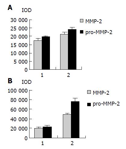
Figure 3 Densitometric intensity of absorbance (IOD) of pro-MMP-2 and MMP-2 activity in colorectal carcinoma tissues.
A: Different lymph node metastases. 1: lymph node negative; 2: lymph node positive; B: Different invasion depths. 1: muscular layer invasion; 2: serous membrane layer or surrounding soft tissue invasion.
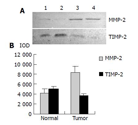
Figure 4 MMP-2 and TIMP-2 expression in normal and colorectal carcinoma tissues.
A: Western blot analysis for MMP-2 and TIMP-2 expression. Lanes 1 and 2: normal tissues; lanes 3 and 4: colorectal carcinoma tissues; B: Densitometric intensity of absorbance (IOD) of MMP-2 and TIMP-2 expression in normal and colorectal carcinoma tissues.
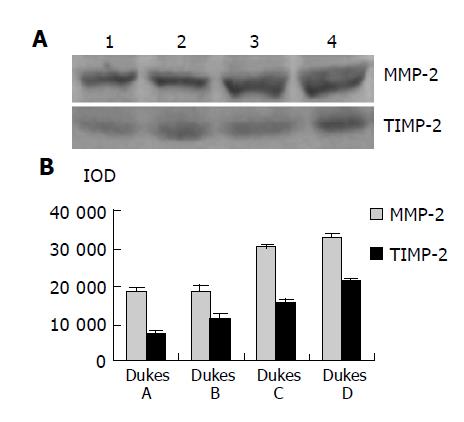
Figure 5 Western blot analysis for MMP-2 and TIMP-2 expression in carcinoma tissues at different Duke’s stages.
Lane 1: Duke’s A stage; lane 2: Duke’s B stage; lane 3: Duke’s C stage; lane 4: Duke’s D stage; B: Densitometric intensity of absorbance (IOD) of MMP-2 and TIMP-2 expression in colorectal carcinoma tissues at different Duke’s stages.
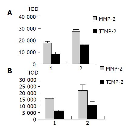
Figure 6 Densitometric intensity of absorbance (IOD) of MMP-2 and TIMP-2 expression in colorectal carcinoma tissues.
A: different lymph node metastases. 1: lymph node negative; 2: lymph node positive; B: Different invasion depths. 1: muscular layer invasion; 2: serous membrane layer or surrounding soft tissue invasion.
Figure 7 MMP-2 and TIMP-2 expression in normal and colorectal carcinoma tissues (SABC ×40).
A: MMP-2 expression in normal colorectal tissue, B: MMP-2 expression in colorectal carcinoma tissue, C: MMP-2 negative control, D: TIMP-2 expression in normal colorectal tissue, E: TIMP-2 expression in colorectal carcinoma tissue, F: TIMP-2 negative control.
- Citation: Li BH, Zhao P, Liu SZ, Yu YM, Han M, Wen JK. Matrix metalloproteinase-2 and tissue inhibitor of metallo-proteinase-2 in colorectal carcinoma invasion and metastasis. World J Gastroenterol 2005; 11(20): 3046-3050
- URL: https://www.wjgnet.com/1007-9327/full/v11/i20/3046.htm
- DOI: https://dx.doi.org/10.3748/wjg.v11.i20.3046


