Copyright
©2005 Baishideng Publishing Group Inc.
World J Gastroenterol. May 28, 2005; 11(20): 3040-3045
Published online May 28, 2005. doi: 10.3748/wjg.v11.i20.3040
Published online May 28, 2005. doi: 10.3748/wjg.v11.i20.3040
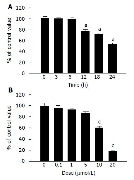
Figure 1 Effect of denbinobin on COLO 205 cell growth.
COLO 205 cells were seeded in a 96-well plate until subconfluence and viable cells were determined by MTT assay. A: Time course study with 10 μmol/L denbinobin in 0.1% DMSO. Two independent experiments were performed in triplicate. aP<0.05 vs time 0. B: Dose-dependent study after treatment of denbinobin for 24 h. Two independent experiments were performed in triplicate. cP<0.05 vs DMSO control.
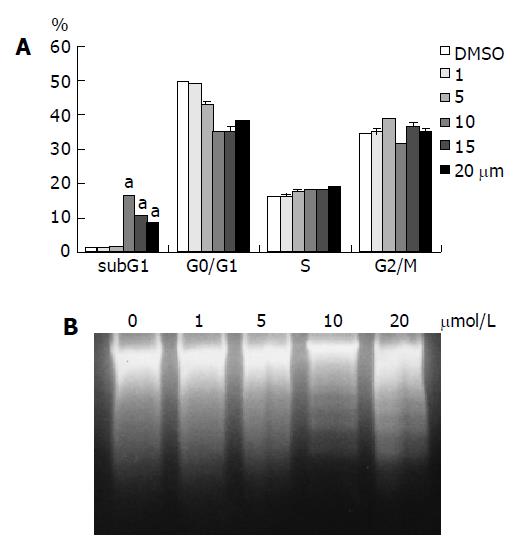
Figure 2 Treatment of COLO 205 cells with denbinobin (10-20 μmol/L) for 24 h induced apoptosis.
A: FACS analysis of DNA content after 24-h incubation in culture medium supplemented with 10% FCS and 0.1% DMSO or 0-20 μmol/L denbinobin in 0.1% DMSO. Percentages of cells in sub-G1, G0/G1, S, and G2/M phases of the cell cycle determined using established CellFIT DNA analysis software were shown. Increased sub-G1 percentage of COLO 205 cells was shown after treatment with 10-20 μmol/L denbinobin. Two independent experiments were performed in triplicate. aP<0.05 vs DMSO control. B: Electrophoresis of genomic DNA from COLO 205 treated with denbinobin. A typical DNA ladder pattern associated with apoptosis was seen at 10-20 μmol/L denbinobin.
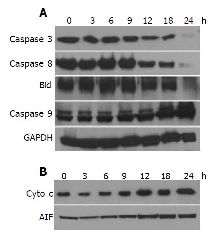
Figure 3 Effect of denbinobin on caspases, Bid protein levels or Cyto c and AIF translocation from mitochondria to cytosol.
The whole cell proteins (A) or cytosolic proteins for Cyto c, AIF (B) were extracted from the cultured COLO 205 cells, which had been grown in 10% FCS and incubated for the indicated times with 0.1% DMSO or 20 μmol/L denbinobin in 0.1% DMSO. After electrophoresis, proteins were transferred onto Immobilon-P membranes, and then probed with proper dilutions of specific antibodies. Membranes were also probed with anti-GAPDH antibody to correct for difference in protein loading. Denbinobin time-dependently induced the activation of caspases and Bid (A), translocation of Cyto c and AIF (B).
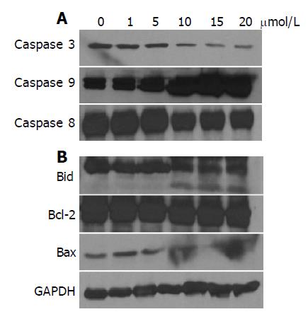
Figure 4 Effect of denbinobin on caspases (A) or Bid, bcl-2 and bax protein levels (B).
The whole cell proteins were extracted from the cultured COLO 205 cells, which had been grown in 10% FCS and incubated for the indicated times with 0.1% DMSO or 20 μmol/L denbinobin in 0.1% DMSO. After electrophoresis, proteins were transferred onto Immobilon-P membranes, and then probed with proper dilutions of specific antibodies. Membranes were also probed with anti-GAPDH antibody to correct for difference in protein loading. Denbinobin dose-dependently induced the activation of caspases (A), Bid protein activation and decreased expression of bax (B).
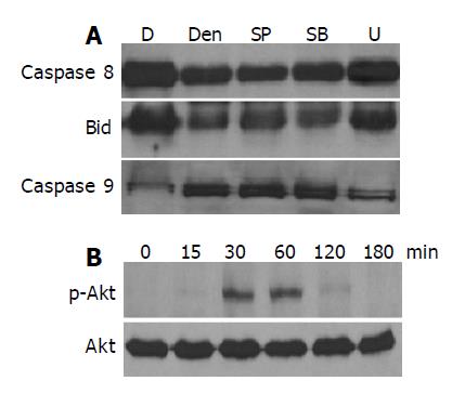
Figure 5 Effect of MAPK and Akt pathways in the denbinobin-induced COLO 205 cells apoptosis.
A: Cells were pretreated with various kinds of MAPKs inhibitors; U10126 (U, 100 nmol/L), SB203580 (SB, 20 μmol/L), or SP600125 (SP, 100 nmol/L) for 1 h. The whole cell proteins were extracted from the cultured COLO 205 cells, which had been grown in 10% FCS and incubated for the indicated times with 0.1% DMSO or 20 μmol/L denbinobin in 0.1% DMSO for 24 h. After electrophoresis, proteins were transferred onto Immobilon-P membranes, and then probed with proper dilutions of specific antibodies; B: Effect of Akt pathway on the denbinobin treatment to COLO 205 cells. Time course study of 20 μmol/L denbinobin-treated COLO 205 cells was used to detect the total Akt proteins and the phorphorylated Akt by Western blot.
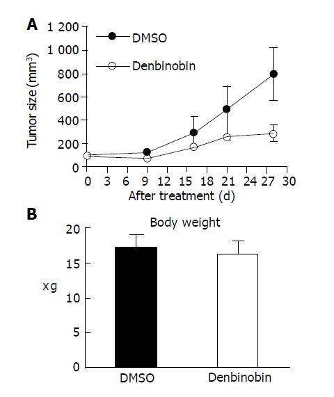
Figure 6 Growth of COLO 205 tumor xenografts in nude mice was suppressed by denbinobin treatment.
Athymic nude mice injected with COLO 205 cells into subcutaneous tissue of inter-scapular area. Once tumor volume reached approximately 200 mm3, the animal received treatment of 50 mg/kg denbinobin or DMSO intraperitoneally thrice per week for 4 wk. (A) Average tumor volume (B) body weight of DMSO-treated (n = 5, filled circles) vs denbinobin-treated (n = 4, open circles) nude mice. Samples were analyzed in each group, and values represent the mean±SE. Comparisons were subjected to linear regression method in (A) and showed significant difference between DMSO vs denbinobin treatment group.
- Citation: Yang KC, Uen YH, Suk FM, Liang YC, Wang YJ, Ho YS, Li IH, Lin SY. Molecular mechanisms of denbinobin-induced anti-tumorigenesis effect in colon cancer cells. World J Gastroenterol 2005; 11(20): 3040-3045
- URL: https://www.wjgnet.com/1007-9327/full/v11/i20/3040.htm
- DOI: https://dx.doi.org/10.3748/wjg.v11.i20.3040


