Copyright
©2005 Baishideng Publishing Group Co.
World J Gastroenterol. Jan 14, 2005; 11(2): 305-307
Published online Jan 14, 2005. doi: 10.3748/wjg.v11.i2.305
Published online Jan 14, 2005. doi: 10.3748/wjg.v11.i2.305
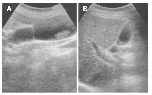
Figure 1 Depicted A polypoid mass in the gallbladder wall depicted by ultrasonography (A).
and A blood clot resolved between 3-7 days after selective arterial embolization (B).
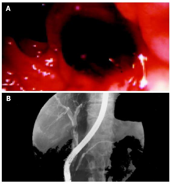
Figure 2 Emanation of fresh blood clots from the ampullar vater (A) and non-homogenous visualization of dilated common bile duct and incomplete visualization of the intrahepatic ducts (B) on endoscopic retrograde cholangiopancreatography.
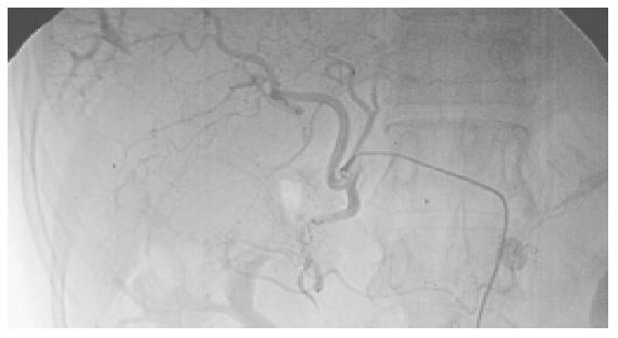
Figure 3 Shunting between a branch of right hepatic artery and right portal vein in segment VII of the liver on arterio-portal fistula on angiography revealing.
- Citation: Lin CL, Chang JJ, Lee TS, Lui KW, Yen CL. Gallbladder polyp as a manifestation of hemobilia caused by arterial-portal fistula after percutaneous liver biopsy: A case report. World J Gastroenterol 2005; 11(2): 305-307
- URL: https://www.wjgnet.com/1007-9327/full/v11/i2/305.htm
- DOI: https://dx.doi.org/10.3748/wjg.v11.i2.305


