Copyright
©2005 Baishideng Publishing Group Co.
World J Gastroenterol. Jan 14, 2005; 11(2): 200-203
Published online Jan 14, 2005. doi: 10.3748/wjg.v11.i2.200
Published online Jan 14, 2005. doi: 10.3748/wjg.v11.i2.200
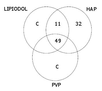
Figure 1 Diagram of nodule detectability by each imaging technique.
Figure 2 A hepatocellular carcinoma nodule identified by Lipiodol CT but not by biphasic MDCT in a 53-year-old man.
A: MDCT scan during the arterial phase shows a 3-cm hypervascular tumor in S5 (arrowhead); B: DSA confirms the MDCT findings (arrowhead); C: Lipiodol CT identifies another tumor in S6 (arrowhead) in addition to S5.
- Citation: Zheng XH, Guan YS, Zhou XP, Huang J, Sun L, Li X, Liu Y. Detection of hypervascular hepatocellular carcinoma: Comparison of multi-detector CT with digital subtraction angiography and Lipiodol CT. World J Gastroenterol 2005; 11(2): 200-203
- URL: https://www.wjgnet.com/1007-9327/full/v11/i2/200.htm
- DOI: https://dx.doi.org/10.3748/wjg.v11.i2.200


