Copyright
©2005 Baishideng Publishing Group Co.
World J Gastroenterol. Jan 14, 2005; 11(2): 182-186
Published online Jan 14, 2005. doi: 10.3748/wjg.v11.i2.182
Published online Jan 14, 2005. doi: 10.3748/wjg.v11.i2.182
Figure 1 Electrophoresis of recombinant plasmid pcDNA3.
1+-mCD40L. Lanes 1 and 5: DNA molecular weight marker; lane 2: The RT-PCR product of mCD40L; lane 3: pcDNA3.1+-mCD40L digested with NheI and EcoRI, pcDNA3.1+ in the upper, full-length mCD40L-cDNA in the lower; and lane 4: pcDNA3.1+-mCD40L not digested with NheI and EcoRI.
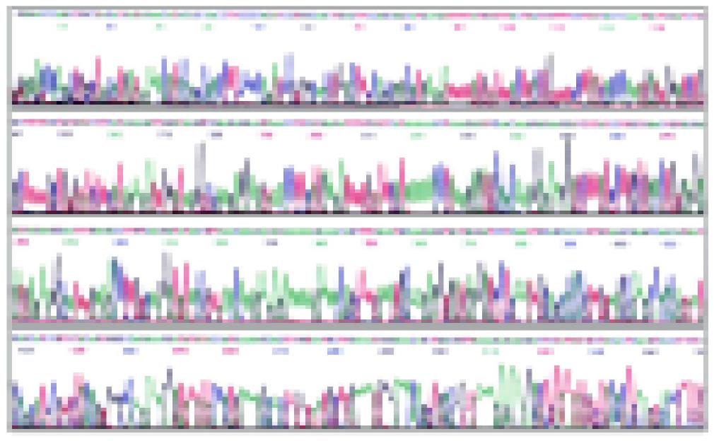
Figure 2 The sequencing map of pcDNA3.
1+-mCD40L with M13F primer.
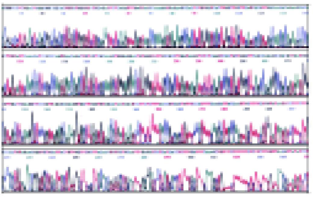
Figure 3 Sequencing map of pcDNA3.
1+-mCD40L with M13R primer.
Figure 4 Nucleotide splicing sequence result of pcDNA3.
1+-mCD40L with DNAstar software analysis.
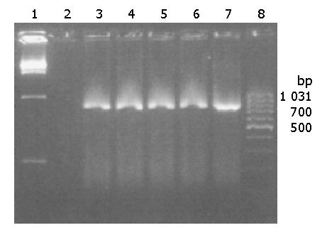
Figure 5 mCD40L gene expression in H22 cells by RT-PCR.
Lanes 1 and 8: marker; Lane 2: negative control; Lanes 3-6: RT-PCR products from transfected H22 cells; Lane 7: RT-PCR product from BALB/C mouse splenocytes.
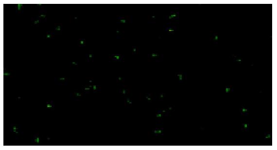
Figure 6 H22 cells transfected with pcDNA3.
1+-mCD40L. The fluorescence staining in the cytoplasm was positive. ×400.
- Citation: Jiang YF, He Y, Gong GZ, Chen J, Yang CY, Xu Y. Construction of recombinant eukaryotic expression plasmid containing murine CD40 ligand gene and its expression in H22 cells. World J Gastroenterol 2005; 11(2): 182-186
- URL: https://www.wjgnet.com/1007-9327/full/v11/i2/182.htm
- DOI: https://dx.doi.org/10.3748/wjg.v11.i2.182


