Copyright
©2005 Baishideng Publishing Group Inc.
World J Gastroenterol. May 21, 2005; 11(19): 2956-2959
Published online May 21, 2005. doi: 10.3748/wjg.v11.i19.2956
Published online May 21, 2005. doi: 10.3748/wjg.v11.i19.2956
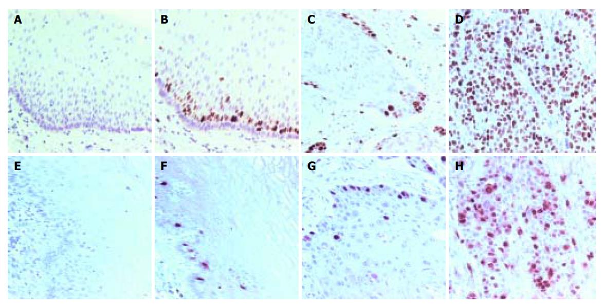
Figure 1 Ki-67 and cyclin A staining patterns in human normal esophageal mucosa and esophageal SCC.
A and E: Negative control in normal esophageal mucosa; B and F: positive nuclear staining in normal esophageal mucosa; C and G: moderately differentiated SCC; D and H: poorly differentiated SCC. Positive nuclear staining was located in base cells in normal mucosa. The diffuse and strong Ki-67 immunostaining in esophageal SCC and the higher SI of the poorly differentiated SCC were found compared with the other carcinomas. Counterstaining with hematoxylin, ×200.
- Citation: Huang JX, Yan W, Song ZX, Qian RY, Chen P, Salminen E, Toppari J. Relationship between proliferative activity of cancer cells and clinicopathological factors in patients with esophageal squamous cell carcinoma. World J Gastroenterol 2005; 11(19): 2956-2959
- URL: https://www.wjgnet.com/1007-9327/full/v11/i19/2956.htm
- DOI: https://dx.doi.org/10.3748/wjg.v11.i19.2956


