Copyright
©2005 Baishideng Publishing Group Inc.
World J Gastroenterol. Mar 21, 2005; 11(11): 1725-1727
Published online Mar 21, 2005. doi: 10.3748/wjg.v11.i11.1725
Published online Mar 21, 2005. doi: 10.3748/wjg.v11.i11.1725
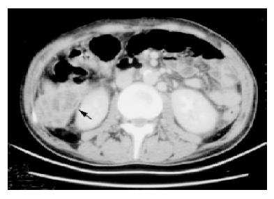
Figure 1 Computed tomography scanning with contrast showed a hypoechoic lesion with irregular and enhanced margin located over segment 6 of liver (arrow), which had ruptured but localized in subhepatic region.
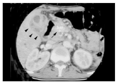
Figure 2 Presence of coarse calcification in the pancreatic head (arrow) indicating presence of chronic pancreatitis.
There is inhomogenous enhancement of the gallbladder wall with pericholecystic fluid, suggesting presence of cholecystitis with empyema (arrowhead).
- Citation: Lai CH, Chen HP, Chen TL, Fung CP, Liu CY, Lee SD. Candidal liver abscesses and cholecystitis in a 37-year-old patient without underlying malignancy. World J Gastroenterol 2005; 11(11): 1725-1727
- URL: https://www.wjgnet.com/1007-9327/full/v11/i11/1725.htm
- DOI: https://dx.doi.org/10.3748/wjg.v11.i11.1725


