Copyright
©The Author(s) 2004.
World J Gastroenterol. Mar 1, 2004; 10(5): 767-768
Published online Mar 1, 2004. doi: 10.3748/wjg.v10.i5.767
Published online Mar 1, 2004. doi: 10.3748/wjg.v10.i5.767
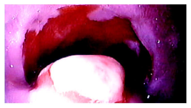
Figure 1 Endoscopic view of inflammatory fibroid polyp (IFP) located in cardia.
An elevated, round, polypoid tumor can be seen.
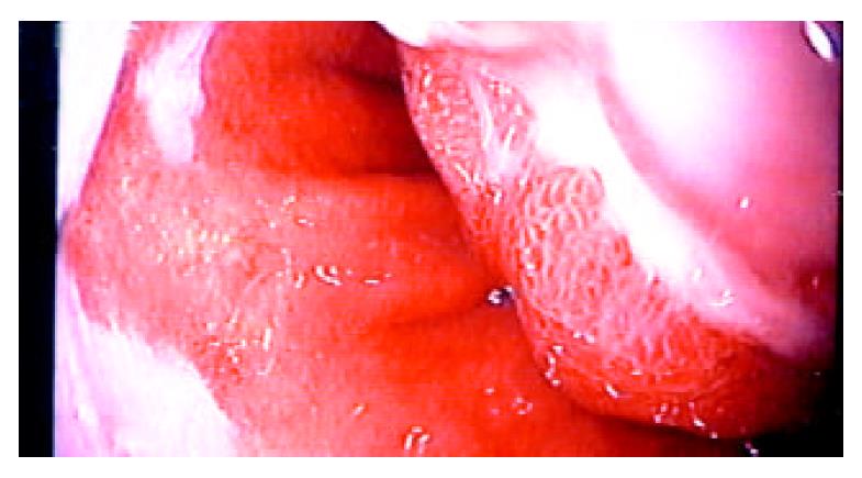
Figure 2 Inflammatory fibroid polyp of cardia.
A small ulcer-ation can be seen in the center of the lesion filled with necrotic tissue.
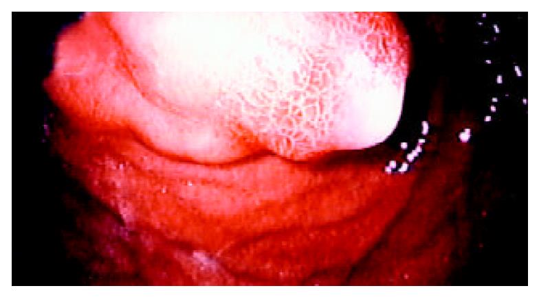
Figure 3 Retroflexion of endoscope.
The tumor is indented into the stomach.
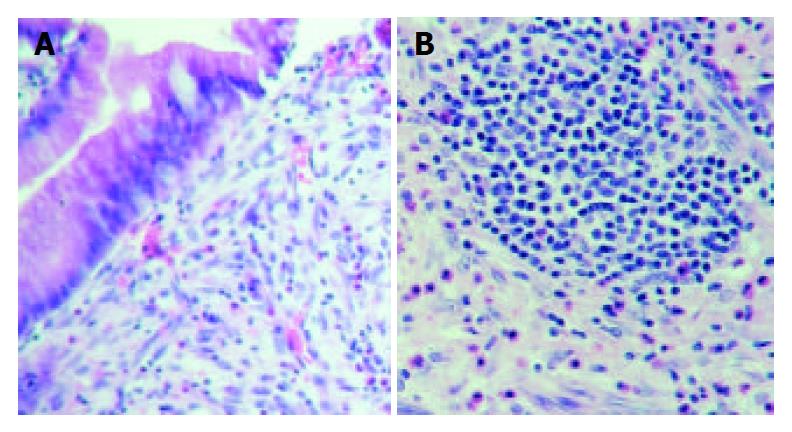
Figure 4 Histologic appearance of primary inflammatory fi-broid polyp of cardia.
A superficially ulcerated tumor can be observed involving cardiac mucosa and submucosa, and con-sists of fibrovascular tissue as well as numerous eosinophils admixed with plasma cells and lymphocytes. (HE; A: magn. ×100; B: magn. ×200).
- Citation: Zinkiewicz K, Zgodziñski W, D¹browski A, Szumi³o J, Æwik G, Wallner G. Recurrent inflammatory fibroid polyp of cardia: A case report. World J Gastroenterol 2004; 10(5): 767-768
- URL: https://www.wjgnet.com/1007-9327/full/v10/i5/767.htm
- DOI: https://dx.doi.org/10.3748/wjg.v10.i5.767


