Copyright
©The Author(s) 2004.
World J Gastroenterol. Mar 1, 2004; 10(5): 660-663
Published online Mar 1, 2004. doi: 10.3748/wjg.v10.i5.660
Published online Mar 1, 2004. doi: 10.3748/wjg.v10.i5.660
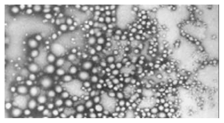
Figure 1 Transmission electron microphotography of TK-PLGA nanoparticles.
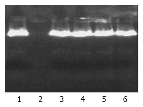
Figure 2 Agarose gel electrophoresis of DNA extracted from nanoparticles after treatment with sonication and Dnase I.
Lane 1 represented untreated control plasmid DNA, lane 2 repre-sented plasmid DNA incubated with Dnase I at 37 °C for 1 h, and lane 3, 4, 5, 6 indicated DNA extracted from PLGA nanoparticles incubated with Dnase I at 37 °C for 0, 4, 8, or 16 h, respectively.
- Citation: He Q, Liu J, Sun X, Zhang ZR. Preparation and characteristics of DNA-nanoparticles targeting to hepatocarcinoma cells. World J Gastroenterol 2004; 10(5): 660-663
- URL: https://www.wjgnet.com/1007-9327/full/v10/i5/660.htm
- DOI: https://dx.doi.org/10.3748/wjg.v10.i5.660


