Copyright
©The Author(s) 2004.
World J Gastroenterol. Mar 1, 2004; 10(5): 638-642
Published online Mar 1, 2004. doi: 10.3748/wjg.v10.i5.638
Published online Mar 1, 2004. doi: 10.3748/wjg.v10.i5.638
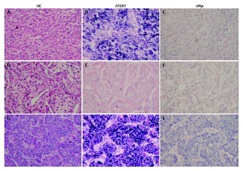
Figure 1 Corresponding distribution of hTERT signals and c-Myc staining in HCC tissue detected by HE staining (A, D, and G); in situ hybridization (hTERT positive, B and H; hTERT negative, E); and immunohistochemistry (c-Myc positive, C and F; c-Myc negative, I).
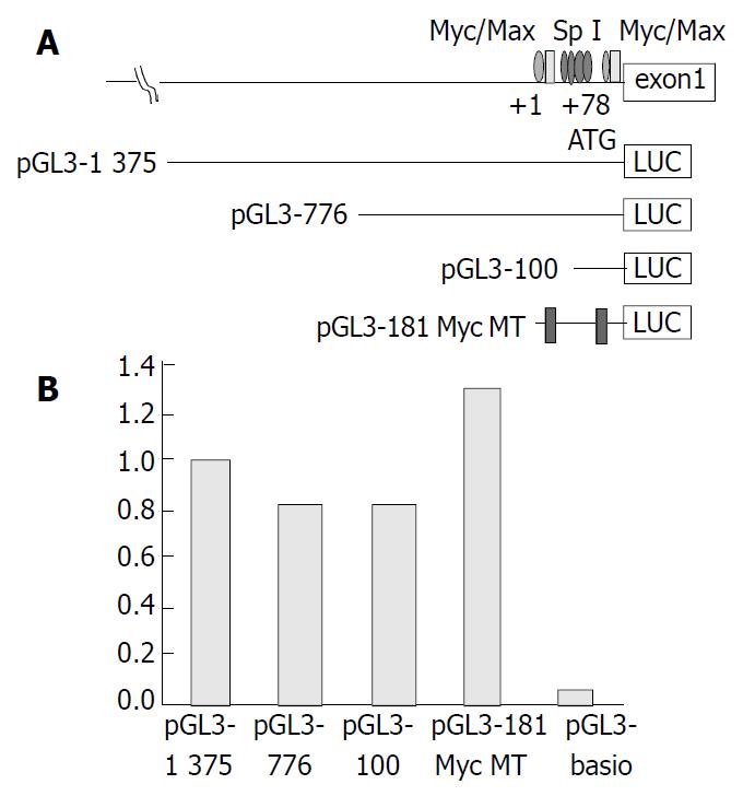
Figure 2 A: Schematic diagram of LUC reporter plasmids.
Pro-moter fragments of decreasing size from the 5’ end (1375 bp, 776 bp, 100 bp and Myc double deletion mutant hTERT-pro-moter reporter plasmids of 181 bp) upstream of the initiating ATG were inserted into luciferase (LUC) reporter vector pGL3-Basic in sense orientation. +1, the transcription start site. Bind-ing sites for c-Myc/Max and Sp1 are shown. B: Transcription activation of hTERT promoter. Data represent normalized rela-tive luciferase fold activity compared with the promoterless pGL3 basic plasmid.
- Citation: Chen CJ, Kyo S, Liu YC, Cheng YL, Hsieh CB, Chan DC, Yu JC, Harn HJ. Modulation of human telomerase reverse transcriptase in hepatocellular carcinoma. World J Gastroenterol 2004; 10(5): 638-642
- URL: https://www.wjgnet.com/1007-9327/full/v10/i5/638.htm
- DOI: https://dx.doi.org/10.3748/wjg.v10.i5.638


