Copyright
©The Author(s) 2004.
World J Gastroenterol. Dec 1, 2004; 10(23): 3511-3513
Published online Dec 1, 2004. doi: 10.3748/wjg.v10.i23.3511
Published online Dec 1, 2004. doi: 10.3748/wjg.v10.i23.3511
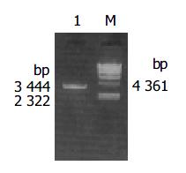
Figure 1 10 g/L agarose gel electrophoresis of cagA DNA fragment amplified by PCR from coccoidal H pylori.
Lane 1. PCR products, Lane M: λ -Hind III DNA marker.
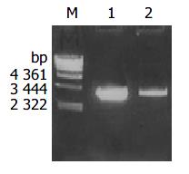
Figure 2 Identification of recombinant vector by PCR.
Lane m: λ -Hind III DNA marker; Lane 1: amplification cagA gene from recombinant pMD-18T-cagA plasmid by PCR; Lane 2: Amplifi-cation cagA gene from coccoid H pylori genome DNA by PCR.
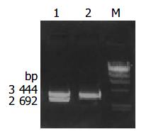
Figure 3 The identification of the pMD-18T-cagA by digestion with restriction endonucleases Lane1: Recombinant pMD-18T-cagA digested by BamH I puls Sac I; Lane 2: Amplification cagA gene from coccoid H pylori genome DNA by PCR; m: λ -Hind III DNA marker.
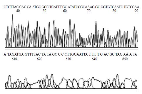
Figure 4 sequencing result of partial cagA gene.
-
Citation: Wang KX, Wang XF. Cloning and sequencing of cagA gene fragment of
Helicobacter pylori with coccoid form. World J Gastroenterol 2004; 10(23): 3511-3513 - URL: https://www.wjgnet.com/1007-9327/full/v10/i23/3511.htm
- DOI: https://dx.doi.org/10.3748/wjg.v10.i23.3511


