Copyright
©The Author(s) 2004.
World J Gastroenterol. Dec 1, 2004; 10(23): 3506-3510
Published online Dec 1, 2004. doi: 10.3748/wjg.v10.i23.3506
Published online Dec 1, 2004. doi: 10.3748/wjg.v10.i23.3506
Figure 1 HDR-192Ir (111-370 GBq) driven to the programmed locations.
A: In the patient who had undergone left-lobe resection due to cholangiocarcinoma, hilar recurrence with bile duct dilation was shown by portal-phase CT scan (arrow). B: The stricture of bile duct was clearly shown by cholangiography during PTCD (arrow). C: An 8 mm×60 mm SMART stent was deployed across the stricture. D: The applicator and sham source were inserted though a 10 F internal-external drainage catheter. The dose distribution curves were obtained from the TPS (arrow).
Figure 2 Cholangiography, CT, or US before and after intraluminal brachytherapy.
A: Bile duct dilation of left lobe caused by cholangiocarcinoma was shown by CT (arrow). B: Cholangiography showed hilar stricture (arrow). C: Intraluminal brachytherapy (fractional dose 7 Gy, total dose 21 Gy) was performed after an 8 mm×60 mm SMART stent insertion. D: CT follow-up 11 mo after intraluminal brachytherapy showed no progress of the tumor and no redilation of the bile duct (arrow).
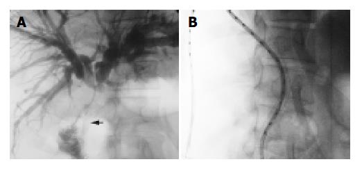
Figure 3 One patient undergoing bilienterostomy and ostomy, which was shown in irradiation region in intraluminal brachy therapy.
A: Cholangiography indicated that jejunum was in-vaded by the malignancy (arrow). B: Jejunum and ostomy were in the irradiation region.
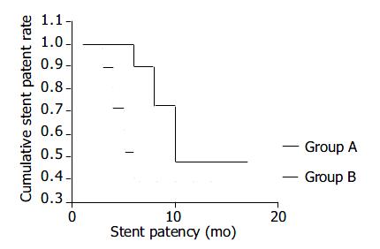
Figure 4 Stent patency of the two groups.
- Citation: Chen Y, Wang XL, Yan ZP, Cheng JM, Wang JH, Gong GQ, Qian S, Luo JJ, Liu QX. HDR-192Ir intraluminal brachytherapy in treatment of malignant obstructive jaundice. World J Gastroenterol 2004; 10(23): 3506-3510
- URL: https://www.wjgnet.com/1007-9327/full/v10/i23/3506.htm
- DOI: https://dx.doi.org/10.3748/wjg.v10.i23.3506


