Copyright
©The Author(s) 2020.
World J Clin Cases. May 26, 2020; 8(10): 1832-1847
Published online May 26, 2020. doi: 10.12998/wjcc.v8.i10.1832
Published online May 26, 2020. doi: 10.12998/wjcc.v8.i10.1832
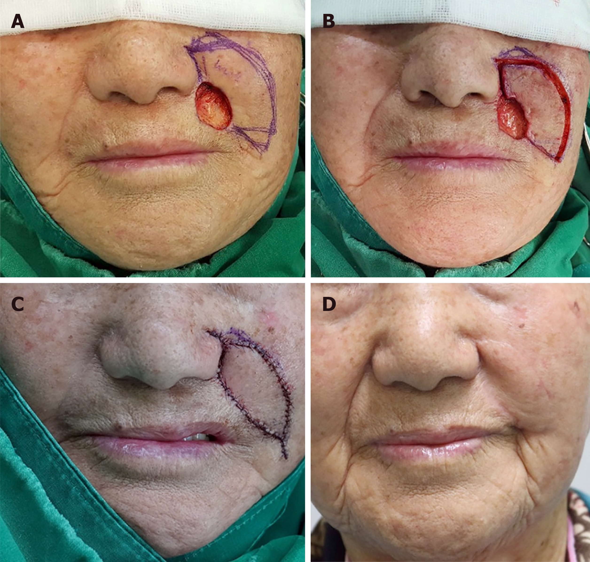
Figure 11 An 82-year-old woman was diagnosed with basal cell carcinoma in the left nasolabial fold area (medial subunit of the cheek unit) by punch biopsy.
A: She underwent a wide excision with a 4-mm safety margin and the final defect was measured to be 2 cm × 3 cm; B, C: We covered the defect with a Type IIA keystone-designed perforator island flap (flap size: 2.5 cm × 5.5 cm) from the upper-lateral side of the defect; D: Postoperative clinical photograph after 6 mo of follow-up. (Reprinted from Yoon et al[1], with permission from Wolters Kluwer).
- Citation: Lim SY, Yoon CS, Lee HG, Kim KN. Keystone design perforator island flap in facial defect reconstruction. World J Clin Cases 2020; 8(10): 1832-1847
- URL: https://www.wjgnet.com/2307-8960/full/v8/i10/1832.htm
- DOI: https://dx.doi.org/10.12998/wjcc.v8.i10.1832


