Copyright
©The Author(s) 2015.
World J Clin Cases. May 16, 2015; 3(5): 405-417
Published online May 16, 2015. doi: 10.12998/wjcc.v3.i5.405
Published online May 16, 2015. doi: 10.12998/wjcc.v3.i5.405
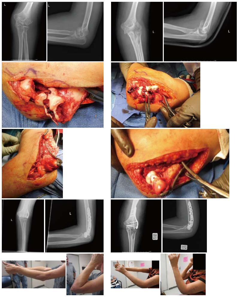
Figure 4 Comparison of outcomes following lateral extensile approach open reduction and internal fixation.
Pictures in the left column represent a 52 years old female who fell from a 2 feet wall. Pictures in the right column represent a 45 years old female who fell from a standing height. As can be seen in the first two rows of pictures in each column, both these patients sustained similar Dubberley type 3b fractures. The third and fourth rows in each column shows that good reduction and fixation was achieved intra-operatively in both patients. The last row in each column show maximum range of motion in each patient. The patient in the left column achieved a reasonable outcome, while the patient in the right column had severe stiffness and range of motion deficit which required an operative contracture release. Both patients underwent open reduction and internal fixation of their fractures using the lateral extensile approach for which the lateral collateral ligament was divided. Secondary to poor results, we have abandoned this method and now universally use the technique shown in Figure 4 or utilize olecranon osteotomy.
- Citation: Yari SS, Bowers NL, Craig MA, Reichel LM. Management of distal humeral coronal shear fractures. World J Clin Cases 2015; 3(5): 405-417
- URL: https://www.wjgnet.com/2307-8960/full/v3/i5/405.htm
- DOI: https://dx.doi.org/10.12998/wjcc.v3.i5.405


