Copyright
©The Author(s) 2024.
World J Clin Cases. Jan 26, 2024; 12(3): 495-502
Published online Jan 26, 2024. doi: 10.12998/wjcc.v12.i3.495
Published online Jan 26, 2024. doi: 10.12998/wjcc.v12.i3.495
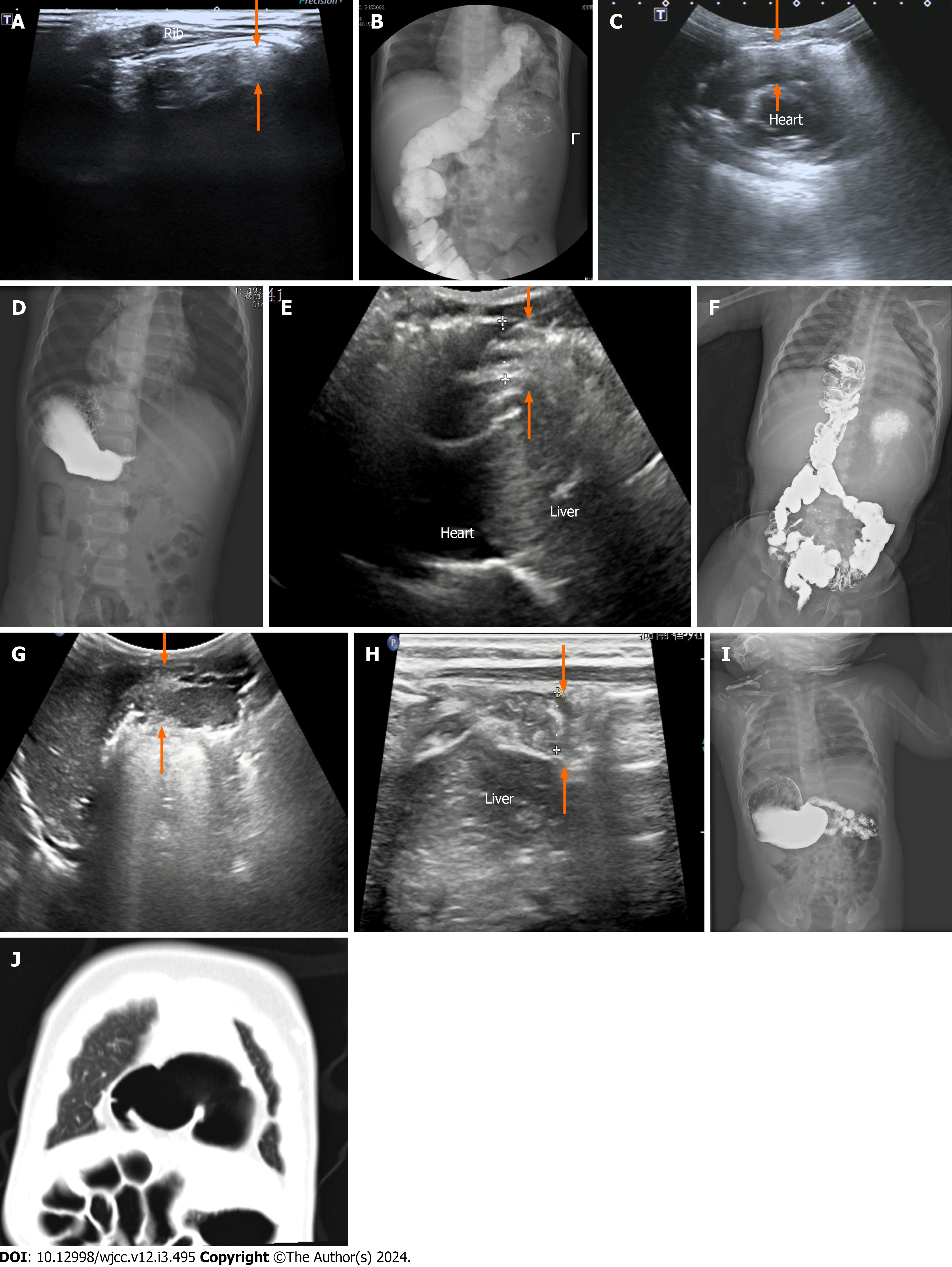
Figure 1 A ultrasound of Morgagni hernia.
A: Case 1: Longitudinal ultrasound (US) scan beside the xiphoid: The intestinal tube and gas echo (arrows) can be seen in left Morgagnian foramina; B: Gastrointestinal imaging (GI) of Morgagni hernia (case 1). Contrast GI showing the herniation of the transverse colon into the left thoracic cavity; C: Case 2: Longitudinal US scan beside the iphoid: Intestinal tube and intestinal gas echo (arrows) between the sternum and the heart; D: GI of Morgagni hernia (case 2). GI suggesting the herniation of the bowel into the right side of the thorax; E: Case 3: Longitudinal US scan beside the xiphoid: Intestine and intestinal gas echoes (arrows) between the sternum and the liver; F: GI of Morgagni hernia (case 3). GI suggesting the herniation of the bowel into the right side of the thorax; G: Case 4: A: Longitudinal US scan of the anterior chest. The echo of intestinal tube and intestinal gas (arrows) can be seen in the right Morgagnian foramina; H: Ultrasound of Morgagni hernia (case 4). US of same patient as in Figure 1G Herniation of the bowel (arrows) into the right side of the thorax; I: GI of Morgagni hernia (case 4). GI showing no obvious abnormality; J: Computed tomography (CT) Morgagni hernia (case 4). Cross-sectional chest CT scan showing the herniation of the intestine into the right thoracic cavity.
- Citation: Shi HQ, Chen WJ, Yin Q, Zhang XH. Ultrasound diagnosis of congenital Morgagni hernias: Ten years of experience at two Chinese centers. World J Clin Cases 2024; 12(3): 495-502
- URL: https://www.wjgnet.com/2307-8960/full/v12/i3/495.htm
- DOI: https://dx.doi.org/10.12998/wjcc.v12.i3.495


