Copyright
©The Author(s) 2023.
World J Clin Cases. Aug 16, 2023; 11(23): 5530-5537
Published online Aug 16, 2023. doi: 10.12998/wjcc.v11.i23.5530
Published online Aug 16, 2023. doi: 10.12998/wjcc.v11.i23.5530
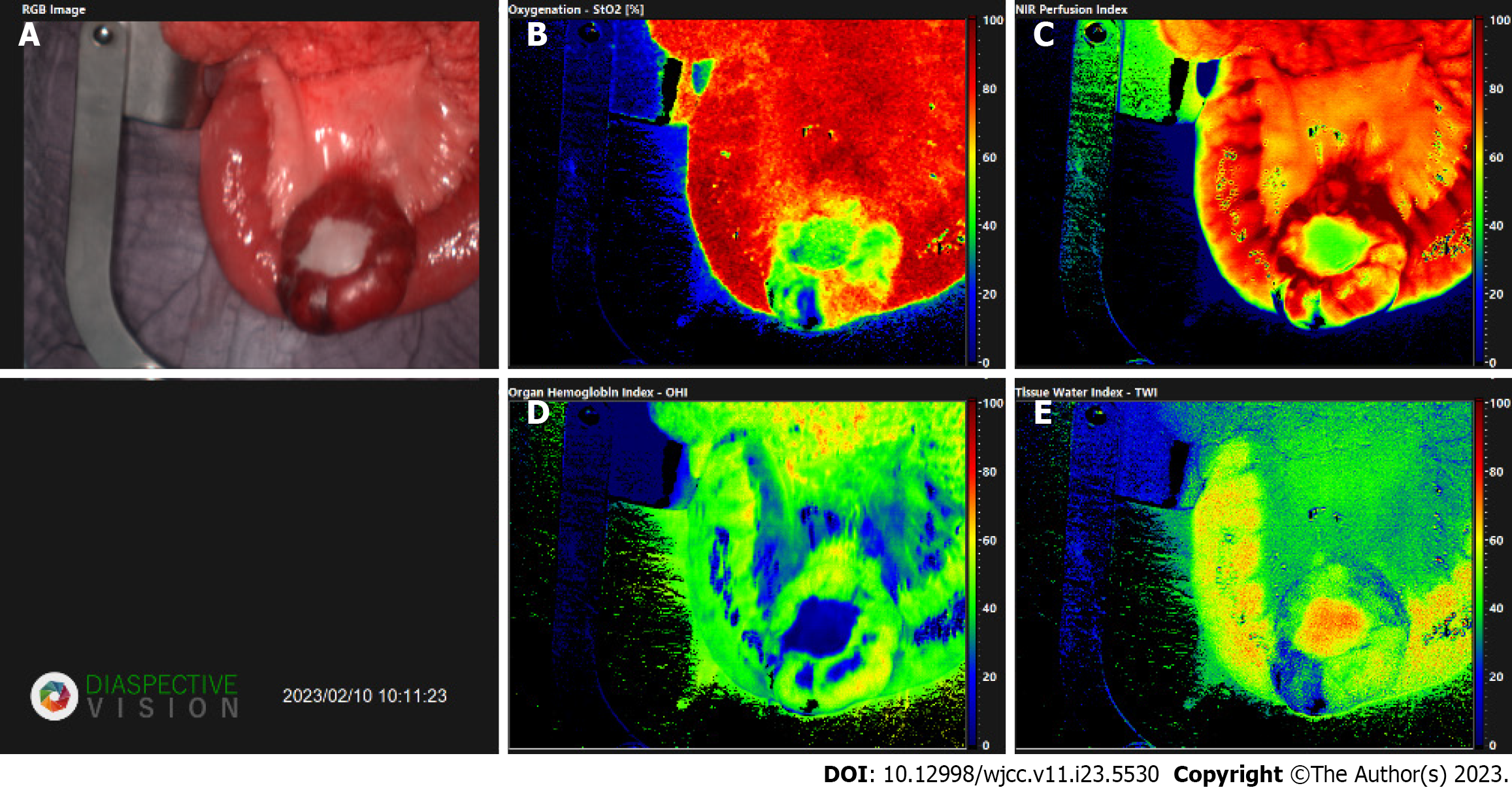
Figure 5 Images acquired via TIVITA® Tissue system (Diaspective Vision GmbH, Am Salzhaff, Germany).
The software provides a red–green–blue image (RGB image) and four false-color images with an effective number of 640 × 480 pixels, which respectively represent tissue oxygenation, near-infrared perfusion index, tissue water index, and organ hemoglobin index[5]. A: Red–green–blue image; B: Oxygenation; C: Near infra-red perfusion index; D: Organ hemoglobin index; E: Tissue- water- index.
- Citation: Wagner T, Mustafov O, Hummels M, Grabenkamp A, Thomas MN, Schiffmann LM, Bruns CJ, Stippel DL, Wahba R. Imaged guided surgery during arteriovenous malformation of gastrointestinal stromal tumor using hyperspectral and indocyanine green visualization techniques: A case report. World J Clin Cases 2023; 11(23): 5530-5537
- URL: https://www.wjgnet.com/2307-8960/full/v11/i23/5530.htm
- DOI: https://dx.doi.org/10.12998/wjcc.v11.i23.5530


