Copyright
©The Author(s) 2022.
World J Clin Cases. Oct 6, 2022; 10(28): 10130-10135
Published online Oct 6, 2022. doi: 10.12998/wjcc.v10.i28.10130
Published online Oct 6, 2022. doi: 10.12998/wjcc.v10.i28.10130
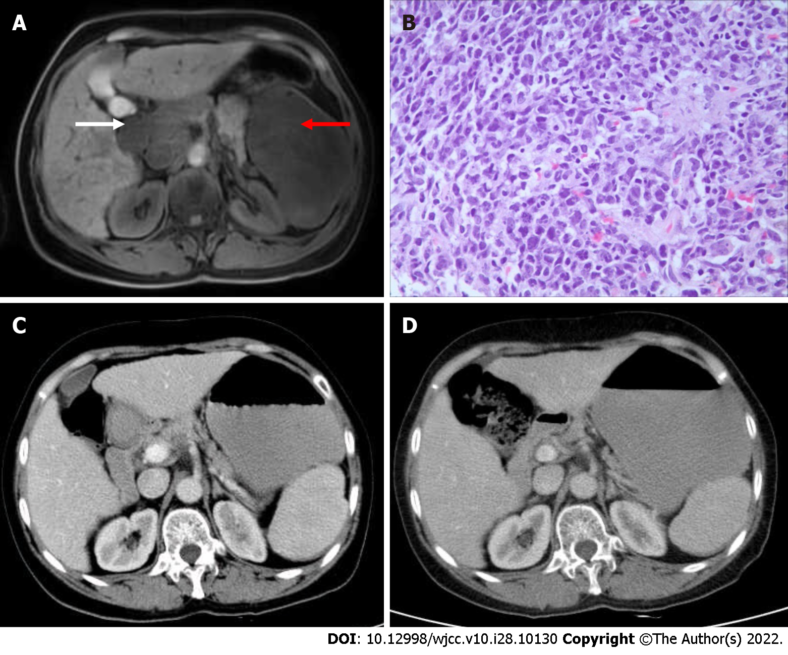
Figure 3 Patient’s magnetic resonance, pathology, and computed tomography images.
A: Axial magnetic resonance imaging showed large lumps with low signals in the spleen (red arrow) on May 22, 2015. Lymph nodes were integrated into a group around the hilum and peritoneum (white arrow); B: Pathological examination of the splenic mass showed a patchy distribution of tumour cells and the disappearance of normal structures (haematoxylin–eosin staining 400 ×); C: Portal phase hepatic enhanced computed tomography (CT) examination after two rounds of R-CHOP treatment on August 19, 2015. Compared with before treatment (Figure 1), the splenic lesions and enlarged lymph nodes in the liver helium and peritoneum had shrunk, and the tumour had disappeared; D: CT scan of the patient at the last follow-up. There was no sign of tumour recurrence at follow-up.
- Citation: Wu FZ, Chen XX, Chen WY, Wu QH, Mao JT, Zhao ZW. Multiple primary malignancies – hepatocellular carcinoma combined with splenic lymphoma: A case report. World J Clin Cases 2022; 10(28): 10130-10135
- URL: https://www.wjgnet.com/2307-8960/full/v10/i28/10130.htm
- DOI: https://dx.doi.org/10.12998/wjcc.v10.i28.10130


