Copyright
©The Author(s) 2022.
World J Clin Cases. Aug 6, 2022; 10(22): 7642-7652
Published online Aug 6, 2022. doi: 10.12998/wjcc.v10.i22.7642
Published online Aug 6, 2022. doi: 10.12998/wjcc.v10.i22.7642
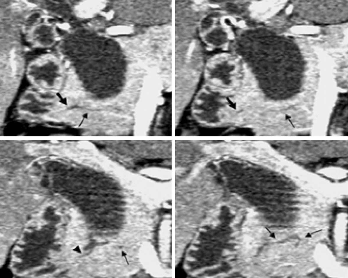
Figure 5 Computed tomography.
Coronal images generated from pancreatic phase scanning show that the pancreatic and biliary ducts join within pancreatic parenchyma. Furthermore, these images make it possible to visualize common channel (arrowhead) and ventral pancreatic duct (thin arrows), which is narrow and tortuous. Thick arrows indicate dorsal pancreatic duct[51]. Citation: Itoh S, Fukushima H, Takada A, Suzuki K, Satake H, Ishigaki T. Assessment of anomalous pancreaticobiliary ductal junction with high-resolution multiplanar reformatted images in MDCT. AJR Am J Roentgenol 2006; 187: 668-675. Copyright © The Authors 2022. Published by American Roentgen Ray Society.
- Citation: Wang JY, Mu PY, Xu YK, Bai YY, Shen DH. Application of imaging techniques in pancreaticobiliary maljunction. World J Clin Cases 2022; 10(22): 7642-7652
- URL: https://www.wjgnet.com/2307-8960/full/v10/i22/7642.htm
- DOI: https://dx.doi.org/10.12998/wjcc.v10.i22.7642


