Copyright
©The Author(s) 2022.
World J Clin Cases. Aug 6, 2022; 10(22): 7642-7652
Published online Aug 6, 2022. doi: 10.12998/wjcc.v10.i22.7642
Published online Aug 6, 2022. doi: 10.12998/wjcc.v10.i22.7642
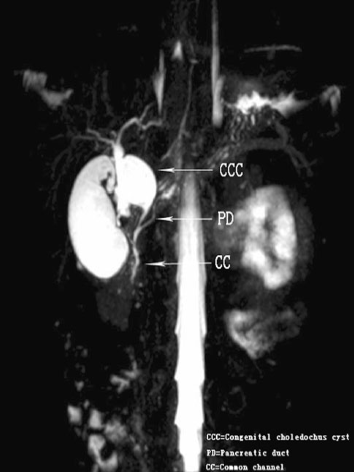
Figure 3 Magnetic resonance cholangiopancreatography.
Coronal 4 mm-thick half fourier acquisition single shot turbo spin echo image shows the pancreatic duct joining the common bile duct outside the duodenal wall[24]. Citation: Guo WL, Huang SG, Wang J, Sheng M, Fang L. Imaging findings in 75 pediatric patients with pancreaticobiliary maljunction: a retrospective case study. Pediatr Surg Int 2012; 28: 983-988. Copyright © The Authors 2022. Published by Springer Nature Switzerland AG.
- Citation: Wang JY, Mu PY, Xu YK, Bai YY, Shen DH. Application of imaging techniques in pancreaticobiliary maljunction. World J Clin Cases 2022; 10(22): 7642-7652
- URL: https://www.wjgnet.com/2307-8960/full/v10/i22/7642.htm
- DOI: https://dx.doi.org/10.12998/wjcc.v10.i22.7642


