Copyright
©The Author(s) 2018.
World J Clin Cases. Jun 16, 2018; 6(6): 121-126
Published online Jun 16, 2018. doi: 10.12998/wjcc.v6.i6.121
Published online Jun 16, 2018. doi: 10.12998/wjcc.v6.i6.121
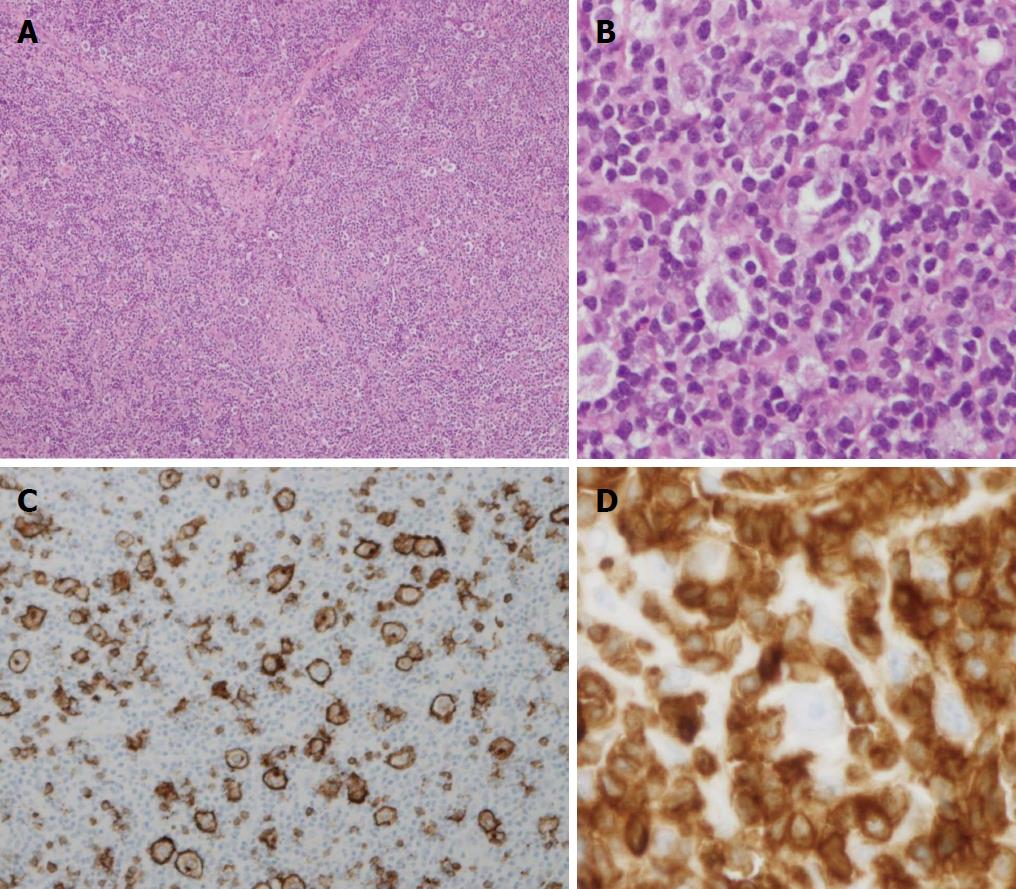
Figure 1 Histopathologic feature.
A: Routine H and E. Scattered large neoplastic cells in background of small lymphocytes and focal sclerosis (40 ×); B: Higher magnification view of neoplastic cells (H and E 200 ×); C: Immunohistochemical stains: Large neoplastic cells marked by CD20 (200 ×); D: One large neoplastic cell surrounded by CD3+ small T-cells, neoplastic cells were negative for CD3 (400 ×).
- Citation: Wei C, Wei C, Alhalabi O, Chen L. T-cell/histiocyte-rich large B-cell lymphoma in a child: A case report and review of literature. World J Clin Cases 2018; 6(6): 121-126
- URL: https://www.wjgnet.com/2307-8960/full/v6/i6/121.htm
- DOI: https://dx.doi.org/10.12998/wjcc.v6.i6.121


