Copyright
©The Author(s) 2024.
World J Clin Cases. May 16, 2024; 12(14): 2389-2395
Published online May 16, 2024. doi: 10.12998/wjcc.v12.i14.2389
Published online May 16, 2024. doi: 10.12998/wjcc.v12.i14.2389
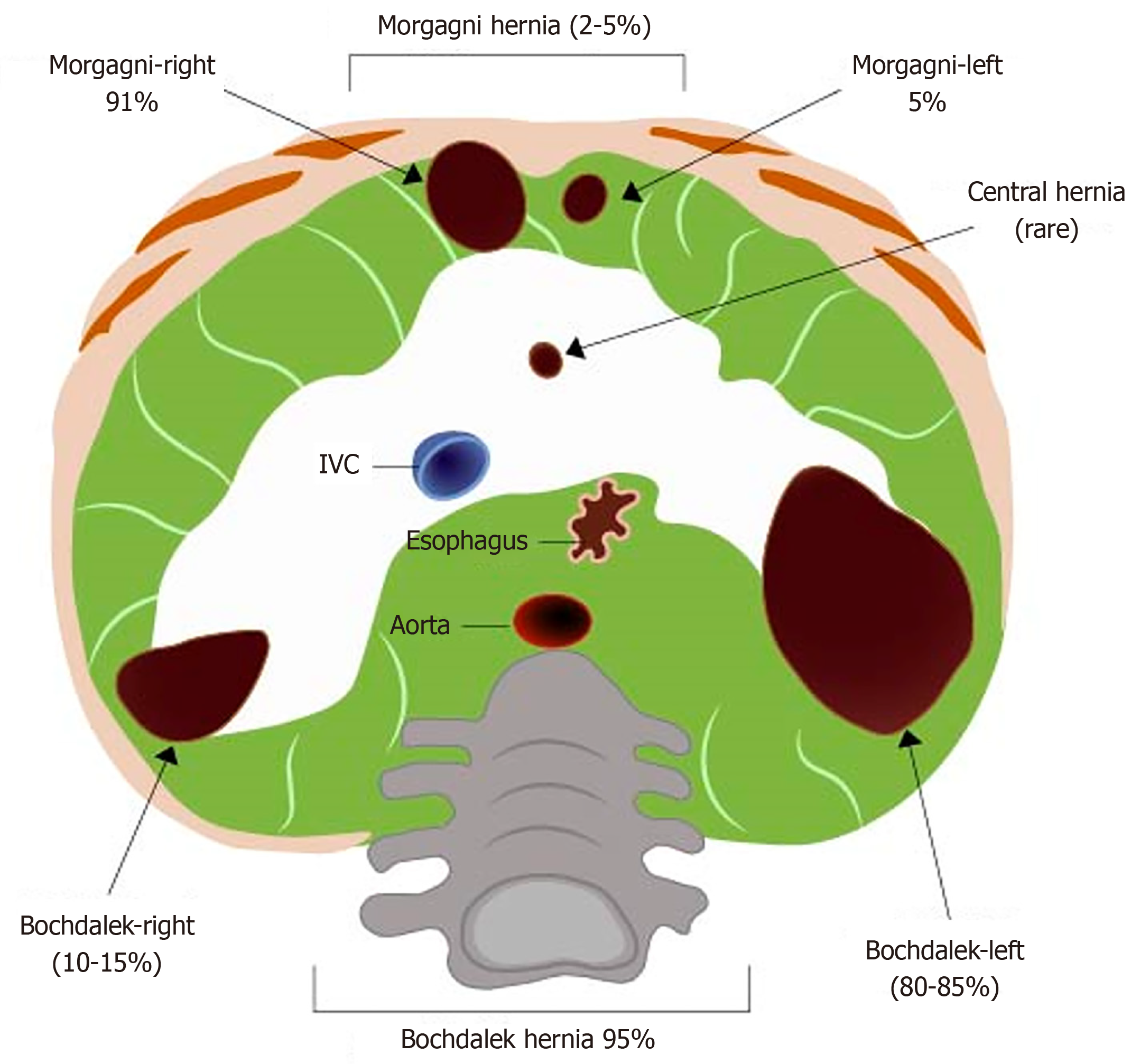
Figure 1 Sites of congenital diaphragmatic hernia formation.
IVC: Inferior vena cava. Modified from Chandrasekharan et al[8].
Figure 2 Colonoscopy appearance of the herniated bowel through Morgagni hernia.
A: Volvulus like appearance; B: Submucosal haemorrhage and evidence of barotrauma.
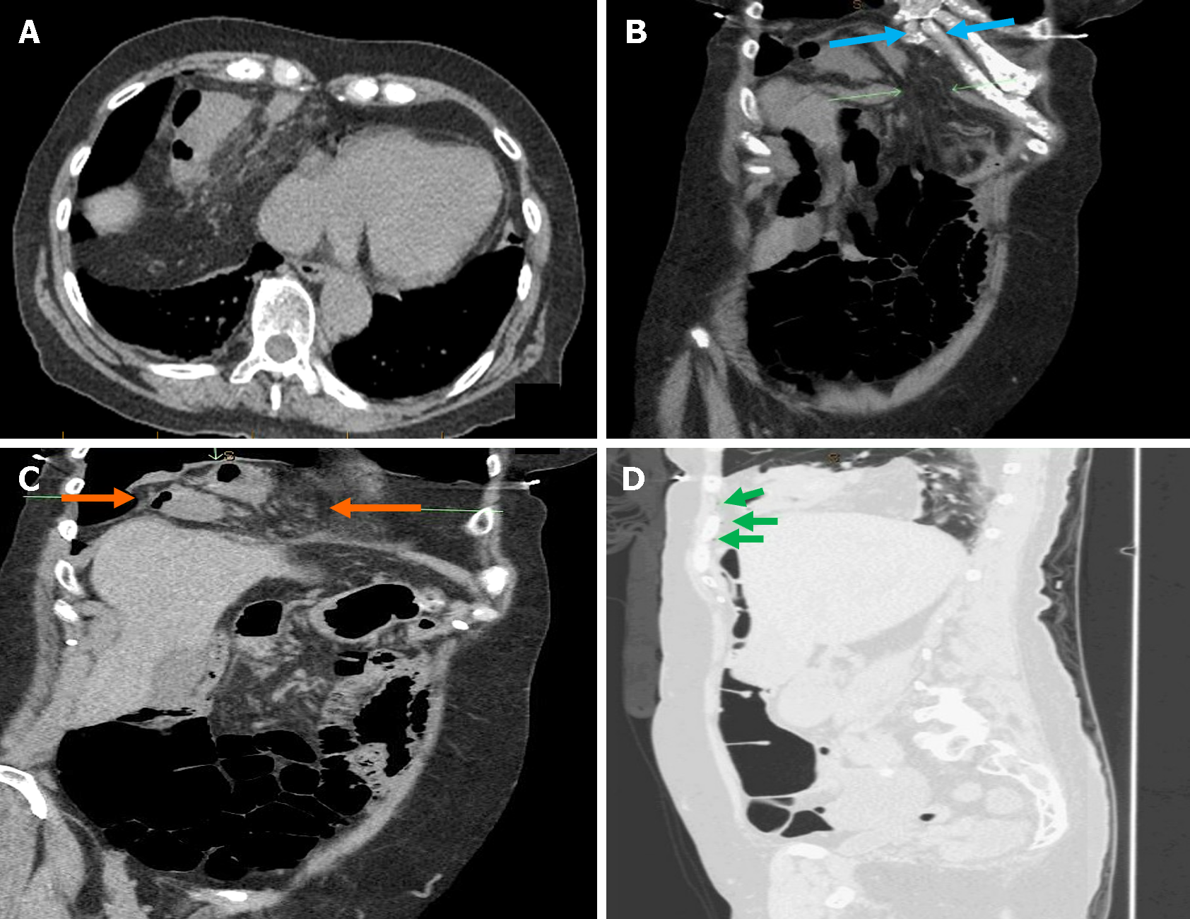
Figure 3 Non-contrast computed tomography scan.
A: Axial view shows incarcerated bowel loops and fat at right paramedian location just above the hemidiaphragm; B and C: Coronal view shows focal defect in the anteromedial aspect of right hemidiaphragm (blue arrows). The hernia sac is seen at right hemidiaphragm and containing bowel loops and mescentric fat (orange arrows); D: Sagittal view in lung window clearly depicts few extraluminal air locules at the non-dependent part secondary to the bowel perforation (green arrows).
- Citation: Al Alawi S, Barkun AN, Najmeh S. Previously undiagnosed Morgagni hernia with bowel perforation detected during repeat screening colonoscopy: A case report. World J Clin Cases 2024; 12(14): 2389-2395
- URL: https://www.wjgnet.com/2307-8960/full/v12/i14/2389.htm
- DOI: https://dx.doi.org/10.12998/wjcc.v12.i14.2389


