Copyright
©The Author(s) 2025.
World J Nephrol. Jun 25, 2025; 14(2): 105374
Published online Jun 25, 2025. doi: 10.5527/wjn.v14.i2.105374
Published online Jun 25, 2025. doi: 10.5527/wjn.v14.i2.105374
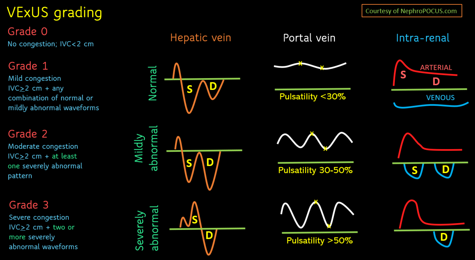
Figure 4 Venous excess ultrasound grading score.
When inferior vena cava has a diameter > 2 cm, hepatic, portal, and rein vein waveforms should be checked. The abnormalities present in these venous Doppler waveforms correlate with the severity of congestion. Hepatic vein Doppler is considered mildly abnormal when the S wave is smaller than the D wave, but still below the baseline; it is considered severely abnormal when the S wave is reversed. Portal vein Doppler is considered mildly abnormal when the pulsatility is 30%-50%, and severely abnormal when it is ≥ 50%. Intrarenal vein Doppler is mildly abnormal when it is pulsatile with distinct S and D components, and severely abnormal when it is monophasic with a D-only pattern. This figure was adapted from NephroPOCUS.com with permission. The corresponding author Koratala A is the owner of the website and copyright holder[60]. See: https://nephropocus.com/about/.
- Citation: Diniz H, Ferreira F, Koratala A. Point-of-care ultrasonography in nephrology: Growing applications, misconceptions and future outlook. World J Nephrol 2025; 14(2): 105374
- URL: https://www.wjgnet.com/2220-6124/full/v14/i2/105374.htm
- DOI: https://dx.doi.org/10.5527/wjn.v14.i2.105374


